//=time() ?>
In this video, Dr. Cohen performs removal of a left medial occipital arteriovenous malformation (#AVM).
See more here:
https://t.co/U09pdR7Nbi
#nsgy #cerebrovascular
Steps for resection of an anterior pontine AVM. Anterolateral view, AVM is medial to the trigeminal nerve, with arterial feeders from the basilar artery, superior cerebellar artery, and anterior inferior cerebellar artery. @mtlawton @BarrowNeuro @BipinChaurasia_ @neuroangio1
Illustration of a glomus spinal AVM. The extrapial portion of the AVM nidus has been resected, leaving the parenchymal portion of the nidus. The glomus spinal AVM has been essentially devascularized and obliterated. By Mark Schornak @BarrowNeuro @neuroangio1 @spinesection
There’s been a few cases of #intracerebral #haemorrhage (ICH) presenting to the #hospital recently. Causes of ICH include hypertension, trauma, AVM or rupture of an aneurysm. In one case I #scrubbed in for an #emergency #crantiomy and the #evacuation of #clot. Image:mayo clinic
Illustration depicting the head positioning, incision, and craniotomy outlines for a typical left frontal AVM. Be sure to note the generous exposure of peri-lesional brain! #brain # neuro #avm #medstudent #neurosurgery
https://t.co/xc50ss56zB
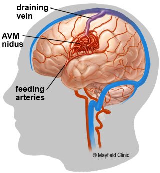
An arteriovenous malformation (AVM) is a tangle of abnormal blood vessels between an artery and a vein. It is susceptible to bleeding leading to a hematoma.
AVM 出品情報
間に合わなかったらゴメンなさい
明日の夜 日本到着予定です。
テイスティングもできていません。
会場限定で店舗でも売りません。
#AVM
Arteriovenous malformation #AVM is an abnormal tangle of blood vessels in the brain or spine https://t.co/49MNm19YXm
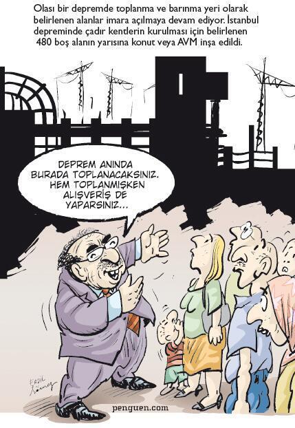
ŞAŞIRDIK MI: Depremde çadır kentlerin kurulması için belirlenen boş alanların yarısı konut veya AVM olmuş