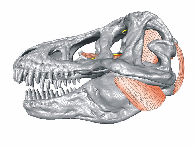
Since then, while the early drafts of the paper loitered on a few desks for a few years #life, some of these ideas leaked into other papers including the 2009 Dino Jaw Muscle anatomy paper and our description of Aegisuchus, which had peculiar vascular scars on its skull roof.
You can listen to me talk about latest discovery on how Alligator and Dinosaur Skulls work here: https://t.co/1JHIn4SxaA
@mizzounews @AnatomyMizzou @MizzouResearch @NSF_BIO
You can find our paper on dinosaur skull roof anatomy and physiology at @anatomyjournal https://t.co/qEa5ZD65C1 https://t.co/hwJPRBT0RW
Next up! Wednesday at #ICVM from @AnatomyMizzou is @kalebsellers presenting work on Crocodilian skull Biomechanics and Evolution in the FEA symposium: 15:30! @nsf_bio @NSF_GEO @MizzouResearch
Be sure to check out Alec Wilken’s talk at #ICVM19 today at 15:30 on his undergrad research project on how Varanid lizard heads work. @NSF_BIO @MizzouUGStudies @MizzouResearch @AnatomyMizzou
Here's a recent article on some of our 3D muscle imaging projects at @Mizzou, @MizzouResearch @AnatomyMizzou @mumedicine. Check it out. https://t.co/IS6T3cz9F7
Imaging is making it easier to see the beauty in anatomy and #sciart, whether its our carpal tunnel (stippling joke!), a xiphisternal flare of a pangolin tongue or the shadowy underpinnings of flight. Sometimes I think its making us lazier, letting machines draw for us.
At some point, I started mixing CT scanned models (plaster Trex) with layered drawings of muscles and nerves, and apparently a stippled quadrate hiding in the back, via Corel. These were never very publishable at the time, so they hang out on my hard drive.
#myfirstspecies was the flat-headed crocodile from northern Africa Aegisuchus witmeri. I stumbled across the specimen in a drawer at the ROM. Named after Larry Witmer.


