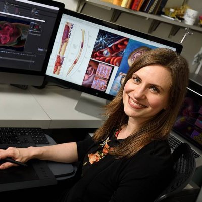
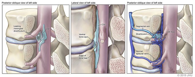
It makes me so happy to sit down at a conference and see my illustrations doing their job!Great talk by Dr. Linda Gray at #SNIS2021 with this illustration of different types of CSF leaks done for Dr. @PeterGKranz!
@SNISinfo
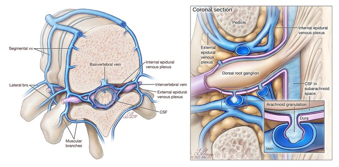
Very excited to have worked with Dr. @PeterGKranz on these illustrations! Normal vertebral venous anatomy, and the relationship between spinal arachnoid proliferations
and the vertebral venous plexus. Brand new paper in @AJR_Radiology: https://t.co/59msCaQj7X
Extremely honored to have worked with Drs. Hans Henkes & Amgad El Mekabaty on this illustration of a pediatric spinal AVF with paraspinal AVM. Full book chapter: https://t.co/7vw0FYeh4c #medicalillustration
Thank you for the prompts @PeterMLawrence1 and @mesabree! Taking advantage of #TBThursday for a blast from the far gone past! #ArtistsOnTwitter
Passing the baton to the talented @srsnyder_biomed @LkethuA @Illustr8Science and @fabiandekok!
#medart #sciart #medicalillustration
In celebration of today's events, a properly treated epidural arteriovenous fistula: red, (off)white, and blue and on the road to recovery! 😁🎉 #medicalillustration
I'll be presenting an overview of my work and the field of medical illustration on Oct. 6, 12pm ET, registration available here:
https://t.co/LKK6YAs9JL Thank you University of MD, Baltimore @umbhshsl for the invitation! @UMMC @UMmedschool @UMBaltimore @AMIdotorg
The 3rd cranial pediatric dural arteriovenous fistula: the rare dural sinus malformation has direct connections between arteries & a sinus with enlarged venous lakes (except for jugular bulb location). For anyone who noticed just 2 in my last post!😁(With Dr. Philippe Gailloud)
Two cranial pediatric dural arteriovenous fistulas: the “adult” type has direct connections between arteries & a venous sinus, the “infantile” type includes the latter with pial arteriovenous shunting, multiple feeders, sinus thrombosis & retrograde flow. For Philippe Gailloud
Good old pen and ink! Overview of the spinal venous system, both intradural and extradural components, which are separate by the dural crossing of the radiculomedullary veins. New article by Dr. Gailloud on the antireflux mechanism located at this point: https://t.co/dA3LH96340
Thoracic vertebral arteries are anastomotic chains similar to cervical vertebral arteries. They frequently provide spinal or bronchial branches, and can be involved in spinal cord ischemia and supply vascular malformations.
Article: https://t.co/apYUM0RUe4


