NephroPOCUSさんのイラストまとめ
@NephroPFollow @NephroPさんをフォローする
フォロー数:307 フォロワー数:87715


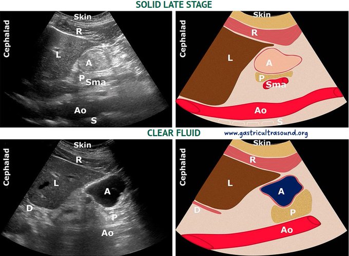
Gastric #POCUS illustrations.
Source: 🔗https://t.co/N17F6DDu2m
A: antrum; Ao: aorta; D: diaphragm; L: liver; P: pancreas; R: rectus abdominis muscle; Sma: superior mesenteric artery
#MedEd #anesthesia #FOAMed


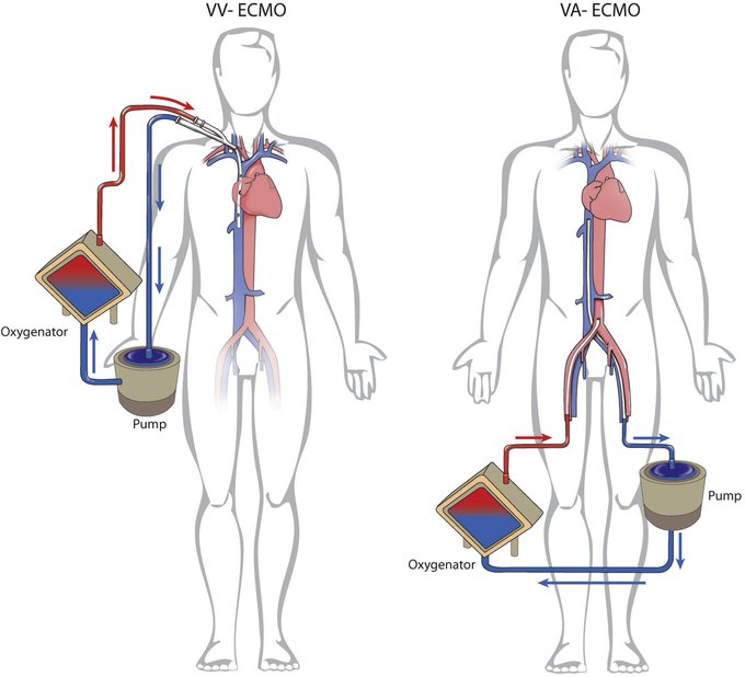
Peripheral VA ECMO cannulation approach: femoral vein (for drainage), (A) femoral, (B) axillary, (C) carotid, artery are used for perfusion.

@NephroMK Not sure why emphysema impedes lateral view because even if the lung is expanded, liver is big enough to provide a window to IVC🤔 If you are using curvilinear probe, try switching to phased array.
Alternatively, can use IJ vein (column height and/or response to Valsalva).

@Rajiv_Sinanan @ThinkingCC @khaycock2 @ArgaizR @katiewiskar @jamiekwillows @ria_dancel Point about arterial interference is more of a caution when rest of the data doesn’t fit. I could be wrong. Whether using subxiphoid or lateral window, location sampled here ‘may’ be subjected to transmitted pulsations.
I remain conservative in this case. Await expert opinion.
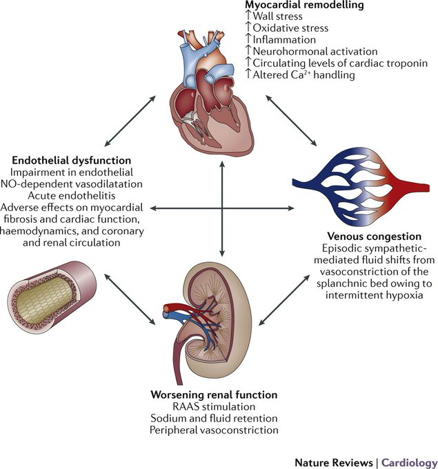
Key pathophysiologic mechanisms underlying acute #heartfailure
#MedEd #Nephrology #cardiorenal
🔗https://t.co/y7JFEU4y3h


Consequences of intra-abdominal hypertension.
🔗https://t.co/Smq3Vw9Mlq
#MedEd #FOAMcc #POCUS