44 件中 41〜44件を表示
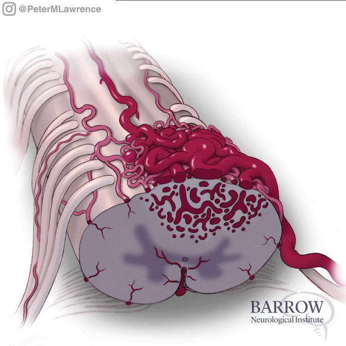
Illustration of a glomus spinal AVM. The extrapial portion of the AVM nidus has been resected, leaving the parenchymal portion of the nidus. The glomus spinal AVM has been essentially devascularized and obliterated. By Mark Schornak @BarrowNeuro @neuroangio1 @spinesection
43
161
Clip from an animation I created a while back that demonstrates a new and improved technique for posterior lumbar interbody fusion (PLIF) with cortical screw fixation by using divergent bilateral interbody grafts. @GlobalSpineJ @BipinChaurasia_ @spinesection
15
71
Can you name these two types of vein of Galen malformations? Created by Mark Schornak, medical illustrator and manager of Neuroscience Publications @BarrowNeuro @BipinChaurasia_ @neuroangio1
27
84


