Here our new publication @Nature #scientificreports Surgical innovation brings improved outcomes and new hope for #pituitary #Acromegaly patients @StanfordNsurg @StanfordMed Great collaborative work by @MohyeldinAhmed @KatznelsonLarry @AcroCommunity https://t.co/2jINX5Ui8k
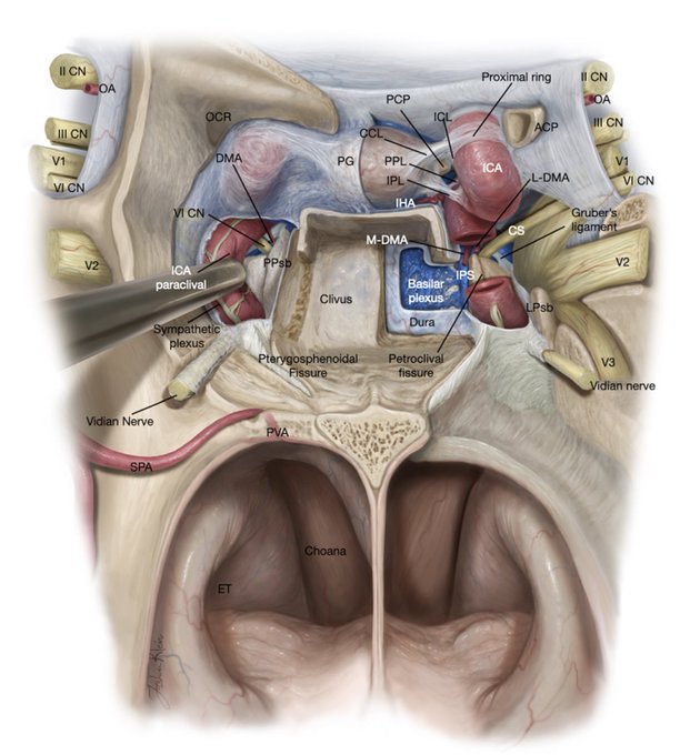
Finding the 6th nerve during #skullbase #surgery may look like a needle in a haystack, but not when you know the key anatomy as published in our latest @TheJNS paper. The spectacular #illustration by Josh Klein @AaronCohenGadol has it all! Our roadmap for #transclival surgery.
✍🏼
SAVE THE DATE! August 10-13, 2022 Innovations in Endoscopic Endonasal Skull Base Surgery. Hands-on dissection course, 3D Lectures & Live Surgery @StanfordMed Master anatomy. Learn new techniques. Improve your practice. Visit Stanford Campus. Limited spots. RSVP.
Let me show you how V2 was finally removed from the cavernous sinus. It literally took a skull base surgeon to do it!! 1st picture, Gray’s Anatomy 1918. 2nd, later editions. 3rd, last 2 editions showing anatomical dissections done at Rhoton’s Lab. Anatomy by surgeons for surgeons https://t.co/xpTh9AtRHR
Thanks for giving us a hand with medial temporal lobe anatomy @RichardRammoMD. During my 2 years in the lab with Prof. Rhoton I studied MTL anatomy with great enthusiasm as it is key to master complex brain tumor surgery. Here the intricate microvascular anatomy of the uncus. https://t.co/7evY8kOJBu


