.さんのイラストまとめ
@sfkhanvisualsFollow @sfkhanvisualsさんをフォローする
フォロー数:333 フォロワー数:39797


It was a mission to correctly identify each structure since the original atlas (atlas of human anatomy and surgery ) doesn’t have labels so really had to put my brain to work 🧠 🫠🫠

Freehand pen and ink study of the internal facial, neck and thoracic anatomy (thought I’d put the rest of my micron pens to use 😛)

A mini infographic on something fascinating I discovered a few weeks ago: the extremely tiny slime molds 🍄🍄🍄


Hello hello #PortfolioDay !! My name is Sana and I’m a medical illustrator and animator 🧠🫁🫀 Currently booking projects for the fall:
✉️: sfkhanvisuals@gmail.com
🌐: https://t.co/ytf7njhauN
🛍: https://t.co/XeqbYyj8mp


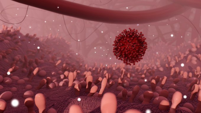
Some of The 3D models from my upcoming masters research project on vaccine mechanisms 👀👀

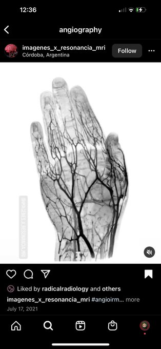
Literally so cool!!! Now I need to find a way to extract such data for a model hmm

OMG thank you @michie_wu for my super cute Avi!!!!!!
She does commissions so check her out ——-> @michie_wu

Combining all of this info, i created this tissue example which gave me a sense of my highlights, shadows, brain and vessel texture in one combined area (sorry for the crummy quality, my scanner is bad)

Another thing to pay attention to is imperfectionsim in realism: not all of the brain gyri ( the puffy parts) are going to be the same size and not all of the blood vessels are going to be symmetrically placed