cranialのTwitterイラスト検索結果。 249 件中 9ページ目


Is the middle cranial fossa a reliable predictor of temporal lobe volume in extant and fossil anthropoids? https://t.co/noeUXJYW2N


In this operative video, Dr. Cohen discusses resection of a craniopharyngioma using an orbitozygomatic craniotomy with a translaminar terminalis approach.
See the pearls here:
https://t.co/vI9bdbZ5sf
#nsgy #tumor #complexcranial
The posterior interhemispheric transcallosal approach exploits a callosotomy just anterior to the splenium that must be preserved. This route also exposes posterior third ventricular and large pineal region tumors with cranial extension.
https://t.co/1zAOV1Crte


Illustration of the formation of intracranial vasa vasorum with atherosclerotic plaque for Dr. Bruce Wasserman. #sciart #medicalillustration https://t.co/MlUxSXDxZA

Patient positioning matters! Here’s a great refresher for incoming #NSGY interns and current residents in this #NeurosurgicalAtlas Ground Rounds: Patient Positioning for Intracranial Surgery with Dr. William Couldwell: https://t.co/lBIDamGpdy @NSTumorSection @cvsection


CRANIAL KINESIS!
- Science vs Care Bears
- T-Rex vs Parrot
- The Science of Scanners' 'splosions!
and MORE at #ZeroPointComedy for free
https://t.co/F1pQdOPW9i


Albrecht Durer (21 May 1471-6 April 1528) "Portrait of a Saint" Provenance from long held German Family Castle (Schloss) Collection from the ancient Habsburg Ducal Line-It possesses all the unique traits- nuances evidenced in his othe "Saints"especially their "Cranial" aspects🧑🎨


#6May #May6 #6Maggio 2020
"Re-examination of the cranial osteology of the Arctic Alaskan hadrosaurine with implications for its taxonomic status"
R. Takasaki et al.
Published: May 6, 2020
PLoS ONE 15(5): e0232410
https://t.co/lhP6qBKZDQ
#fossils #fossilfriday #paleontology


"새로운 형태의 뇌 자기 자극, 우울증에 90% 효과"
https://t.co/RyIOply4tD
자기로 뇌 심부를 자극하는 '경두개 자기 자극'(TMS: Transcranial Magnetic Stimulation)의 방법을 개선하면 우울증 치료 효과를 90%까지 크게 높일 수 있다는 연구 결과가 나왔습니다.


Meanwhile back in coal country, Trump supporters already frustrated with diminished cranial capacity ... are completely confused.


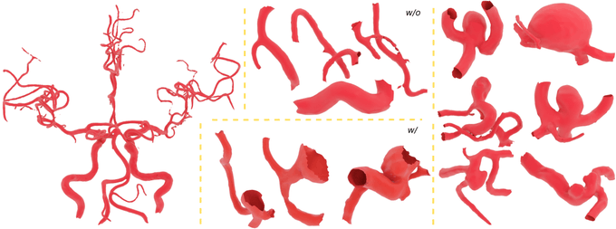

IntrA: 3D Intracranial Aneurysm Dataset for Deep Learning. https://t.co/Eyb6z2mArd


*Must-read* review - multidisciplinary management of intracranial metastases. https://t.co/Q8wk2RUEHY
Excellent! (We expect no less from @DrJohnSuh @ClevelandClinic and my friend Eric Chang @keckmedusc .) #radonc #neurorad #neurosurgery @NeurosurgeryUSC @USCneurorads

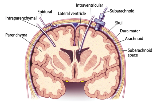
35. There are other methods to monitor ICP in different intracranial locations. These include an extraventricular drain (EVD) or intraparenchymal catheter. These are illustrated below ! #BBTBI


#Brain imprints on cranial bones from great #apes & #humans refute the long-held notion that the human pattern of brain asymmetry is unique. New paper in @ScienceAdvances by @MPI_EVA_Leipzig researcher Simon Neubauer et al. More: https://t.co/lbr6MDb8wh & https://t.co/jiT7pOn102

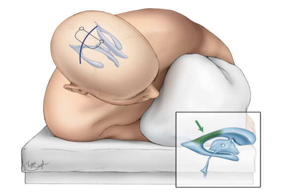
During transcranial resection of tuberculum sella meningiomas, it's crucial to preserve the small branching vessels originating from the superior hypophyseal artery complex & the medial aspect of the internal carotid artery supplying the optic nerve/chiasm
https://t.co/vyQXmbNxZ3
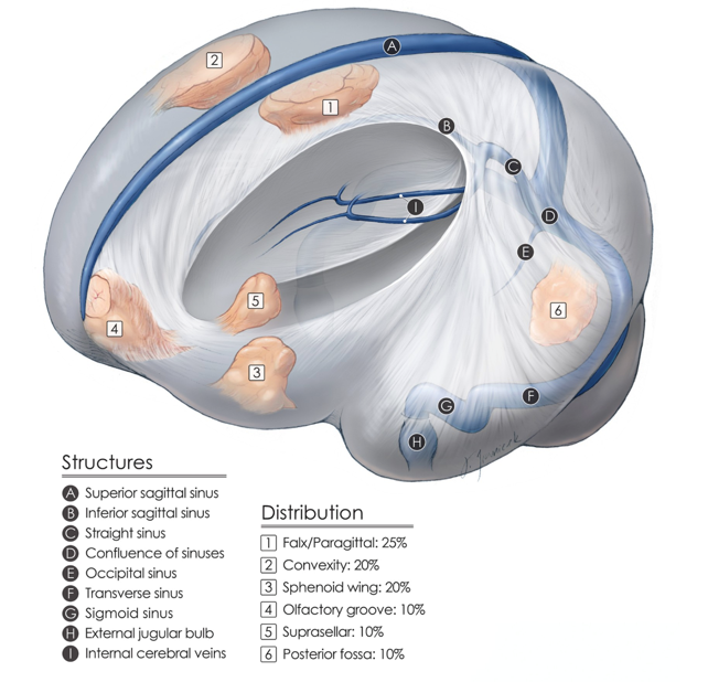
Here, we see the relative distribution of the most common intracranial meningiomas. Resection of these benign tumors is associated with good patient outcomes and therefore is satisfying for neurosurgeons.
#nsgy #tumors #meningioma
https://t.co/Ldg5aBh08O


Detailed information on "Postcranial anatomy and osteoderm histology of Riojasuchus tenuisceps and a phylogenetic update on Ornithosuchidae (Archosauria, Pseudosuchia)" by Belén von Baczko et al. - https://t.co/U5itWr8MAl #paleontology #rioja #argentina #crocodiles #cocodrilos #c


Realityfade bands love post.
🇩🇪Exorcised Gods (Germany)
https://t.co/FdJn9pSyFz
🇬🇧Visions Of Disfigurement (UK)
https://t.co/ol0CFIntVx
🇬🇧Cranial Contamination (UK)
https://t.co/BhTXKSwkf7
🇭🇰🇹🇼 Facelift Deformation (Hong-Kong/Taiwan)
https://t.co/QuQoDFGAGF


Pterional craniotomy-a highly flexible skull base approach that affords excellent exposure of the anterior cranial fossa, the circle of Willis, and the interpeduncular region. @AaronCohenGadol summarizes an efficient execution of this route & its expansion https://t.co/NoRlEe5cqx