NeurosurgeryのTwitterイラスト検索結果。 164 件中 5ページ目


Temporalis muscle (yellow) being affixed to a plate as part of a cranioplasty - the surgical repair of a skull defect from a previous operation/injury. Both the plate and re-attaching the muscle help to restore the natural contour of the skull. #neurosurgery #MedTwitter #anatomy


A dark and stormy look at #biologicalmarkers in the #blood that can lead to earlier #Alzheimer
# PatientEducationillustration # Biologyillustration #Neuroscienceillustration #Neurosurgeryillustration #medicalillustration
By © Lisa A. Clark @lisaannclark
https://t.co/SLjZf8Focy


One of the key investigations carried out in neurology and neurosurgery is the lumbar puncture. This article by @Kiren_Parkash demonstrates the indications for the procedure and how it is performed! A very insightful article for patients & professionals
https://t.co/X6M8oqAUDR


It's #sciartportfolioweek Today I'm sharing some of my favorite neurosurgical illustrations from the last few years. For more of my work, check out https://t.co/EbbrwRthBe #medicalillustration #sciart #neurosurgery @GNSIorg @SupportBarrow


This unique and novel illustration with its layered format provides a road map for surgery within the periclival region. #neurosurgery
See more here:
https://t.co/BfezRCFXqG


Diagrams done for the brilliant @chrishoney - first physician to recognize and treat hemi-laryngopharyngeal spasm (HeLPS syndrome). @GNSIorg #sciartportfolioweek #medicalart #medicalillustration #cranialnerves #neurosurgery

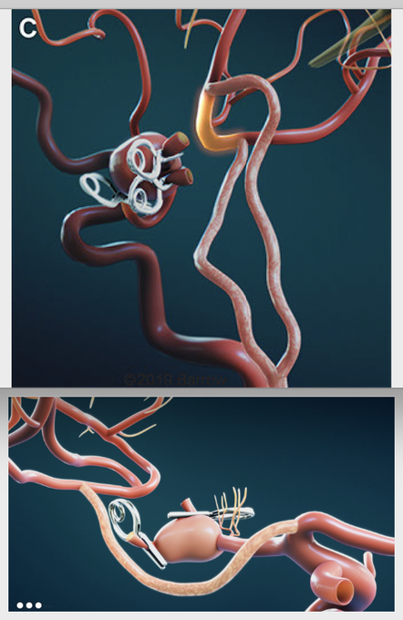
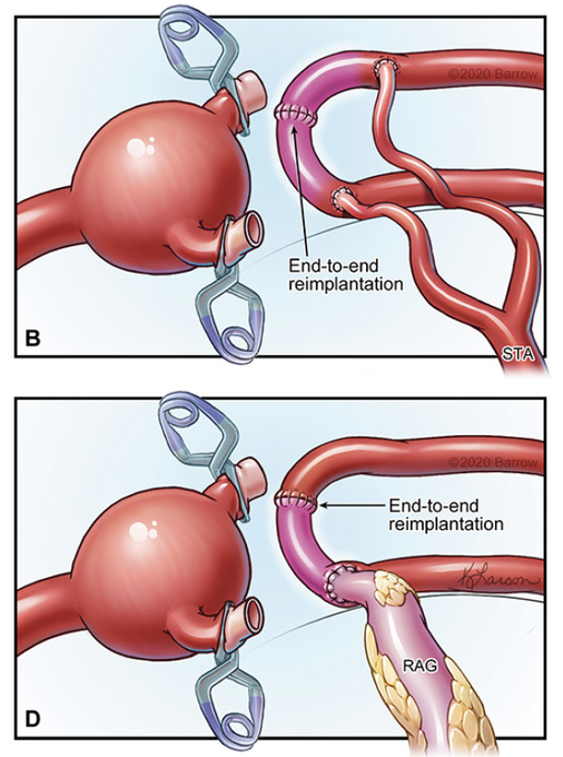
Some say open vascular neurosurgery is fully matured, stagnant. Middle communicating artery exemplifies why that’s not true. It demonstrates 4th generation bypass concept, novel end-to-end reimplantation of M2 trunks, innovation. We are the authors of our future - keep evolving!


Cranium #metastasis illustration for a case report...
Professional illustrations from medical professionals.
#medicalillustration #digitalart #medical #illustration #neurosurgery #medicalart #drawingdoctors #tumour #brain #BrainTumorThursday #sciart

Craniopharyngiomas can have suprasellar components with cystic expansion. Illustrated here is a retrochiasmatic craniopharyngioma, with endoscopic visualization of the tuberculum sellae and anterior wall of the sella outlined in blue. #neurosurgery #tumor
https://t.co/fuJYioaPK2

Kocher’s point maximizes chances of safe placement of an external ventricular drain. A key step is to drill the burr hole and place the catheter perpendicular to the skull, with the catheter aimed toward the foramen of Monro.
#neurosurgery #neuroanatomy
https://t.co/dU3x8QyDy3


For all my #neurosurgery colleagues, the first print in my neuro series, “Clipped Aneurysm,” is now available in all sizes on Etsy https://t.co/Vtrf1BWeWs
How cool is this fenestrated clip?!Stay tuned for more soon!
#MedTwitter #AbstractAnatomy #SurgeryArt #TheSassySurgeon

The basic principles of the anterior interhemispheric transcallosal approach are illustrated. The reach of this approach is shaded in green (inset image). #nsgy #neurosurgery #tumor https://t.co/1zAOV1Crte

Many of the arteries supplying the insula originate from the M2 segment. Long M2 perforators can travel through tumors, but often supply the corona radiata and thus must be preserved during resection of insular lesions. #neurosurgery #brain #neuroanatomy
https://t.co/WqpYe5I1eh

Deep venous anatomy is key in the management of pineal region tumors. Illustrated here is the location of one such tumor through the occipital transtentorial route. #neurosurgery #brain #tumor #nsgy
https://t.co/QlSSjafpvq


Cool suture retraction technique applied to EC-IC #bypass surgery. Similar to @MKBaskayaMD's for trans-sulcal approach (I've used twice before with some <partial> success). Bypass: https://t.co/oB9oOabuFg @WorldNeurosurg
#cerebrovascular #microneurosurgery @cvsection


Painting for a #Neurosurgery trigeminal neuralgia project @IGL_Berlin @vajkoczy

Temporal horn AVMs are technically challenging to tackle because the nidus is covering the feeding vessels emerging from the anterior choroidal artery within the choroidal fissure. #nsgy #neurosurgery #cerebrovascular
Learn more here:
https://t.co/cn2k9dzhwt

For ventricular body AVMs that extend lateral to the midline, a contralateral transcallosal route is favored to minimize ipsilateral hemispheric retraction. #nsgy #neurosurgery #cerebrovascular
Learn more here:
https://t.co/cn2k9dzhwt
During a transsylvian amygdalohippocampectomy, incising the temporal stem allows for access to the temporal horn and serves to create a corridor through which the amygdala and hippocampus can be removed. #neurosurgery #brain #neuroanatomy #nsgy
https://t.co/q4fP0e4Ukc
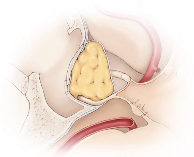
Continuing on with our theme from last week of #skullbase reconstruction: Abdominal or thigh fat can be buttressed with a bony septum or prosthesis to keep the fat in place and reconstruct the sella floor. #nsgy #neurosurgery #medtwitter
https://t.co/ZSbv8Gt1uM