DermpathのTwitterイラスト検索結果。 17 件


Ceruminous gland adenoma: external ear canal, can be painful & cause hearing impairment🔬 Well defined with apocrine inner lining and outer myoepithelial layer. No overt cytologic atypia #Dermpath #Pathology #Dermatology


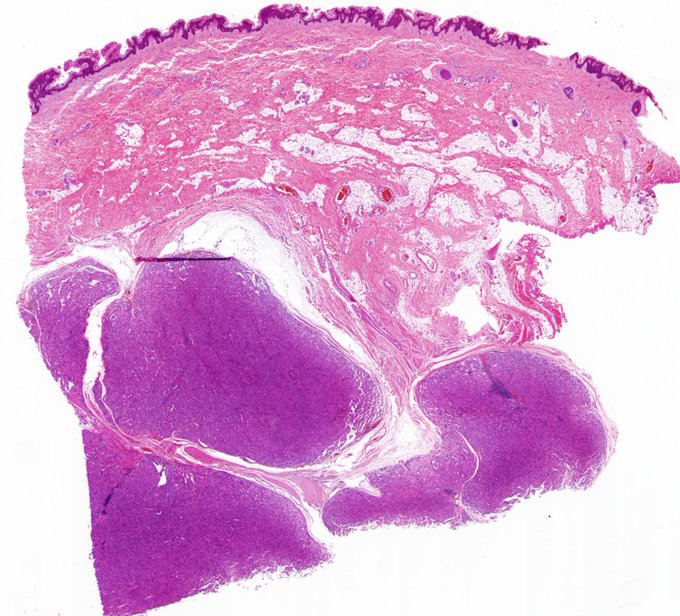
Peri-orbital nodule , adult
#Dermpath #Breastpath #pathtwitter #pathresidents


9 year old girl with a lesion on her lower central back. you diagnostic impressions? #dermpath #pediapath #PathTwitter #coolPathcases
@alegru18 @BattistellaMD @VHNguyenMD @PIorfino @TristanRutland7 @david_terrano @ChristineJKoMD @redsnapperpath @RyanneMDMBA @SGottesmanMD


Carcinoma cuniculatum:
🔬rare type of well-diff SCC which shouldn't be confused with verrucous ca
🐇forms cystically dilated sinuses "the burrows of a rabbit warren"
🦶usually develops on soles
A while ago we proposed its criteria in AJD: 10.1097/DAD.0000000000001457 #dermpath


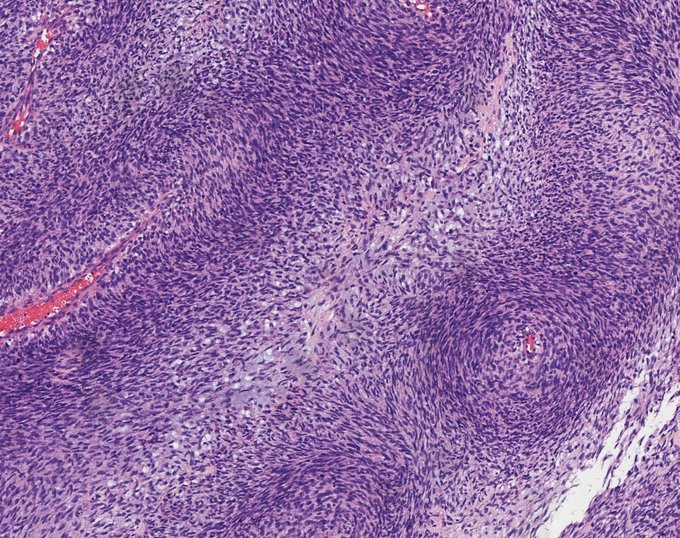
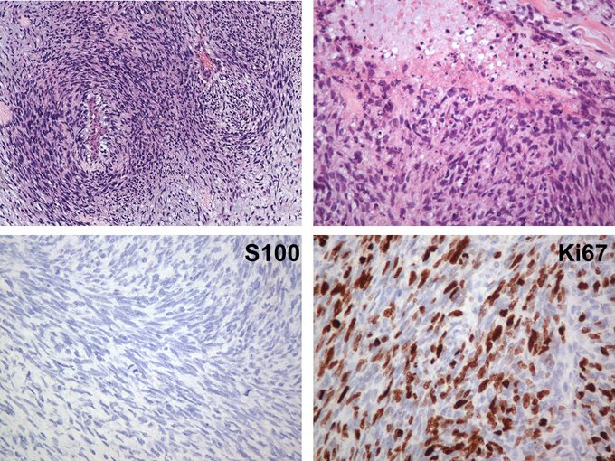
Look at this high-grade cutaneous MPNST! #dermpath #pathology


Thursday #dermpath special. 😆🔬
I could not let this go without sharing its morphologic beauty.
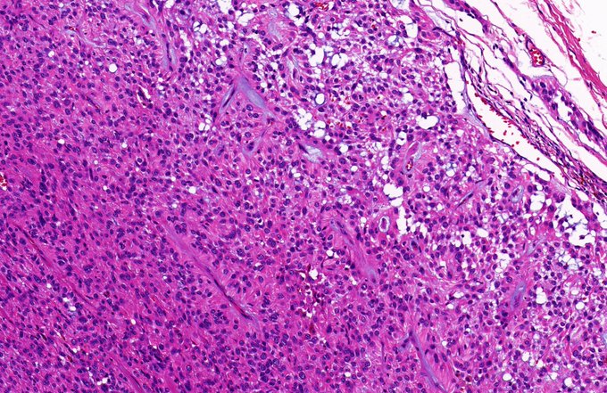
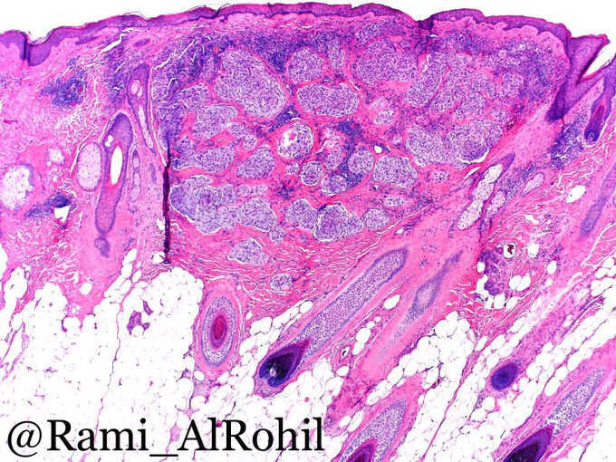
Sebaceoma


20 yo woman. Thumb lesion. A perfect example! What is the diagnosis? Answer: https://t.co/ap5qa7YGWm. Video on how to tell it apart from a potential mimic: https://t.co/VgWjuYhRxV. #pathology #pathologists #pathTwitter #dermpath #dermatology #dermatologia #dermtwitter


He’s back! #PoirotForever Biopsy from the chest of a 60+ year old woman. What do you think is going on here? #dermpath #dermatology #pathology #SilviPearls


36M Hx: lesion left temporal scalp ?BCC/??other. Any guesses?
#pathology #dermpath #histopathology #PathTwitter


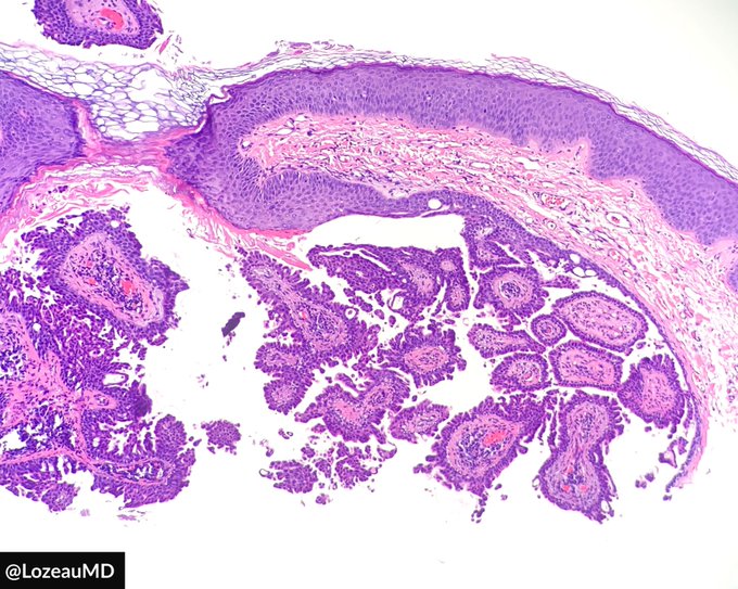

Cystic syringocystadenoma papilliferum! #dermpath #pathology #dermatology #dermtwitter


Happy Saturday! Show a really “not much” case, but turns out again be “very much” and serious. I saw these lesions daily, most are just solar lentigo or macular SK. But you have to be careful and use IHC generously. 66 cheek lentigo.#Dermpath #dermatology #pathology


Think about foreign body (splinter in this case) when there’s extensive epidermal necrosis and intense acute inflammation #Pathology #dermatopathology #Dermpath


Cellular neurothekeoma: solitary lesion, young age group, mostly females on the head&neck. Multilobular pattern in a fibrotic/hyalinized stroma, lobules composed of round cells with vesicular chromatin arranged in concentric nests that swirl w/ some mucin #Pathology #Dermpath

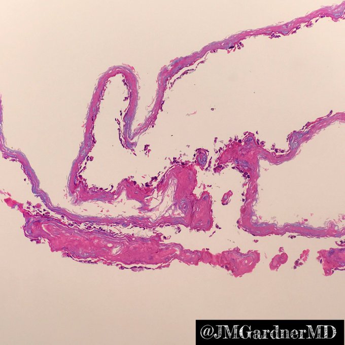

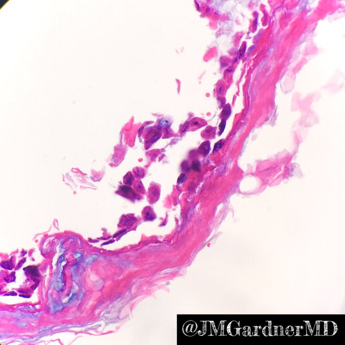
Diagnosis: Staph scalded skin syndrome. SSSS (not SJS/TEN). Detached stratum corneum w free floating acantholytic keratinocytes (key to dx!). If corneum missing look around slide to try to find it. No inflammation or bacteria (toxin mediated). #dermpath #Pathology #dermatology
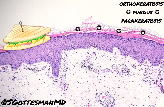
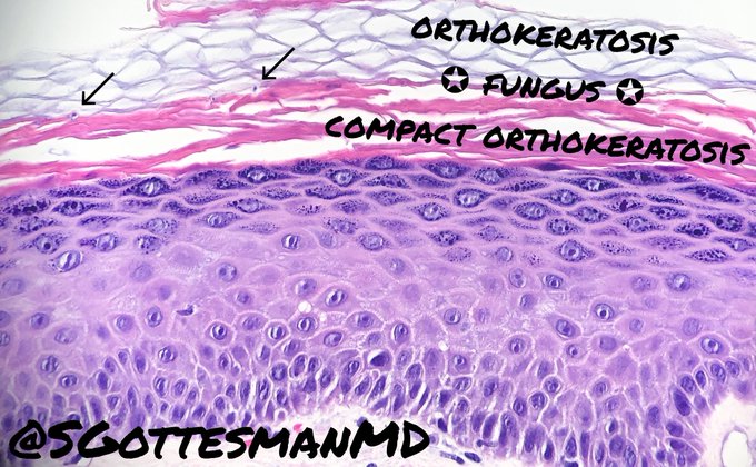
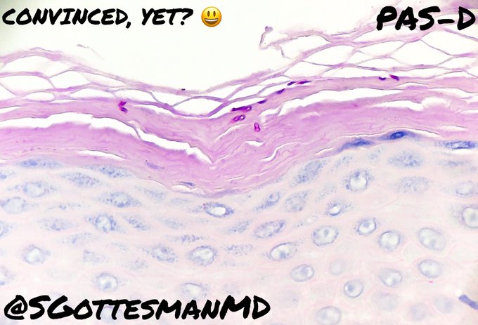
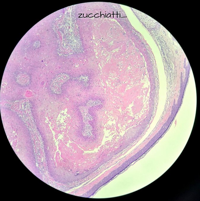
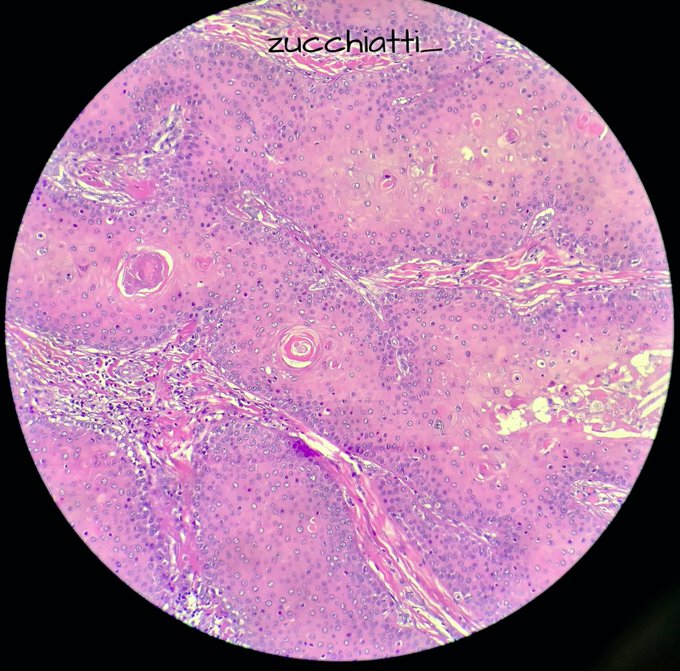
Gottlieb’s sandwich sign: Fungal hyphae are “sandwiched” between two zones of cornified cells: orthokeratosis on top & parakeratosis or compact orthokeratosis below. Can be used as a clue for low power dx of dermatophyte infection. #dermpath #dermatology #SilviPearls #pathology


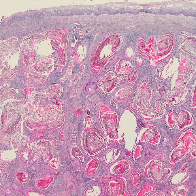
✔ this is a proliferating trichilemmal cyst:
📍proliferation of squamous cystic areas in dermis, lobular architecture.
📍stratified squamous epithelium with abrupt/trichilemmal keratinization.
📍rupture frequent.
📍malignant transformation is rare.
📍DDx with SCC #dermpath