ProteinImagerのTwitterイラスト検索結果。 11 件


Crystal structure of a borosin methyltransferase from Mycena rosella (PDB code: 7TWL) #scivis #sciart #molecularart #proteinimager
https://t.co/xaSWCWE2aU

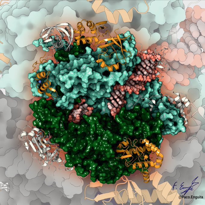
CryoEM structure of the Salmonella typhimurium RuvAB branch migration motor in complex to a Holliday DNA junction. My tribute to the amazing work from @MarlovitsLab (PDB code: 7PBU) #scivis #proteinimager #molecularart #sciart
https://t.co/AUIIkufD5P

Personal interpretation of a recent cryoEM structure of the Escherichia coli retron-Ec86 (RT-msDNA-RNA) obtained at 3.2 angstrom (PDB code: 7V9U) #sciart #scivis #proteinimager #cryoem #molecularart
https://t.co/tKuZ0dsxai

Crystal structure of the Bacillus cereus NaK channel (PDB code: 7OPH) #scivis #sciart #molecularart #proteinimager
https://t.co/zHn1xoHiQE

Structure of the vaccinia virus transcription initiation complex determined by CryoEM (PDB code: 7AOZ)
New #rendering by @fenguita (Paco.Enguita on Instagram) made with #ProteInimager.
Here is the 3D view: https://t.co/ES1UBdZoYi
#molecularart

New #rendering by @fenguita (Paco.Enguita on Instagram) made with #ProteInimager.
Here is the 3D view: https://t.co/iA2pNLByqy
Crystal structure of a telomeric RNA G-quadruplex complexed with an acridine-based ligand.
#molecularart #immolecular

New #rendering by @fenguita (Paco.Enguita on Instagram) made with #proteinimager.
Here you have an unpublished crystal structure of SmAP protein from the bacterium Methanococcus vannielii in complex with UDP (PDB code: 5MKN)
Explore in 3D at https://t.co/mMXqAi3D44
#cryoEM 2.4 Å resolution structure of the Light-Harvesting 2 complex from the marine purple bacterium Mch. purpuratum on @ScienceAdvances. Rendered by #ProteinImager user @valentina_VP17. PDBid 6ZXA


Teaser Alert! In the next release of #ProteinImager there will be the ‘quantized’ style option! For those days when you feel just a little crazier than usual🤪
#sciart #visualization

PhD student @valentina_VP17 having fun with a structure solved in 1994 (PDBid 1eri): EcoRI endonuclease binding #DNA, BFF of all molecular biologists out there.
Made w/ #ProteinImager https://t.co/fA8dSbfmI5

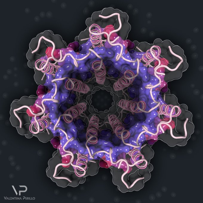
High-resolution structure of DHBV capsid-like particles (CLPs) determined by electron cryo-microscopy (PDB code: 6YGH).
.
Rendered by @fenguita (@Paco.Enguita on Instagram) with #proteinimager . Here the link to 3D structure https://t.co/zMSTStGGAC
.
#sciart #biology #science