anatomy4sculptorsのTwitterイラスト検索結果。 26 件
The thorax and the rest of the skeleton stay the same, regardless of the body type. A person can gain or lose weight and muscle mass, but the bony framework will remain unchanged. #anatomy #references #art #anatomy4sculptors
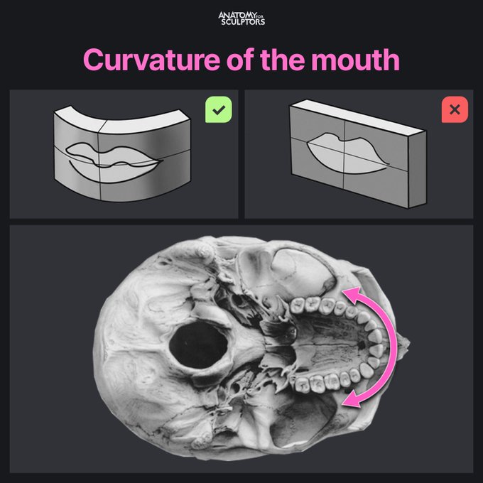
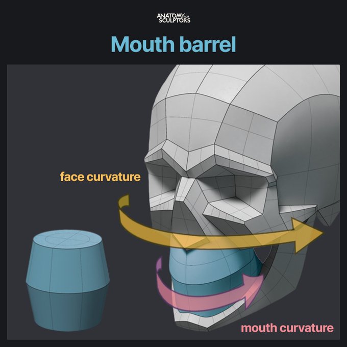
The mouth area has a sharper curvature than the rest of the face. Tip for artists: recognize the shape of the mouth barrel and organize it around a smaller radius than the face. #anatomy #references #art #anatomy4sculptors
The first dorsal interosseous is a muscle mass located between the thumb and the index finger metacarpals on the backside of the hand. Squeeze your thumb to your index finger and see for yourself: this muscle mass pops out and creates an egg-shaped bulge. #anatomy4sculptors
GM! Saturday mornign is anatomy practice, so went back to this dude from last week and refined him a little bit. Using Anatomy4Sculptors ecorche as reference.
Almost all of the palm is covered with fat except a triangular fat-free spot in the middle. Thenar eminence, Hypothenar eminence, Interdigital area, and the fingers all feature fat pads, which explain the hand's imprint, as seen in the picture. #anatomy4sculptors
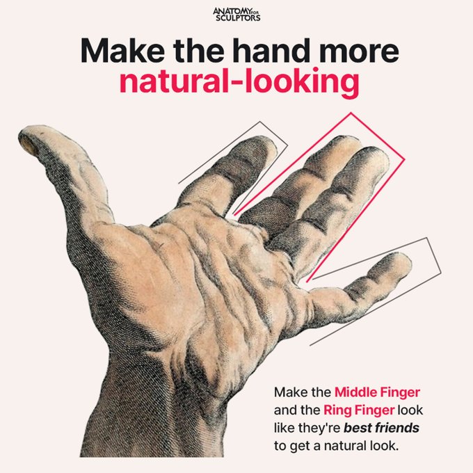
Make the middle and ring fingers stick together to get a more natural look for the hand. #anatomy4sculptors
Read our latest blog on creating a realistic human 3D model! Our senior 3D artist Jēkabs Jaunarājs shares his methods for authoring skin textures: https://t.co/U4O9QamTCt
Check out other blogs in the series to learn about creating eyes, hair, and teeth. #anatomy4sculptors
Male buttocks from simple shapes to complex anatomy! Start with two short cylinders and work towards complex organic forms. #anatomy4sculptors
The Gluteus maximus, gluteus minimus, and tensor fascia latae are the gluteal muscles responsible for the shape of the hips and buttocks. Observe the picture with the muscle overlay and try to notice these muscles in the live model photo! #anatomy4sculptors
During scapular protraction, the scapula and all the muscles attached to it move forward along with the arm. The scapula glides freely along the surface of the thorax: the curved shape of the scapula matches the big spherical egg that the thorax is. #anatomy4sculptors
A sequence of steps from a complex block-out of the head and neck toward the simple bases for the sketching phase. Two commonly used head and neck bases are the egghead and the helmet head.
View the detailed block-out 3D model here: https://t.co/dI9BycSzP0 #anatomy4sculptors
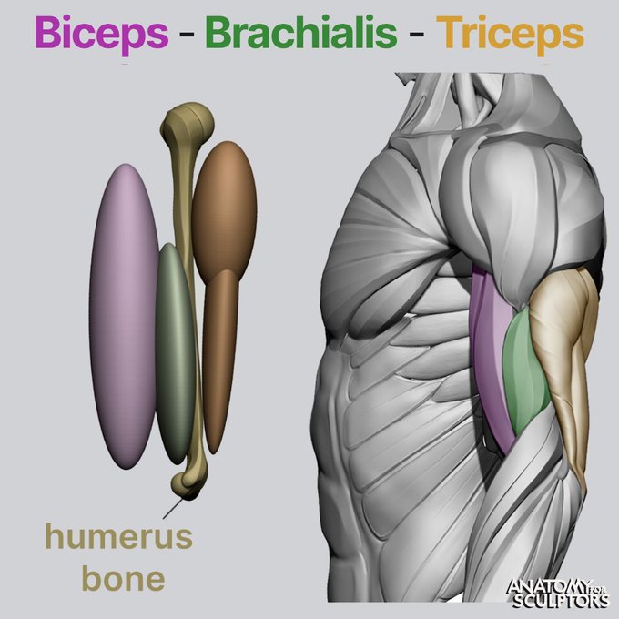
When thinking about the upper arm, the biceps and triceps are the first muscles that come to mind. It is equally important to remember the brachialis muscle, which is located right underneath the biceps, thus creating the base for the flexors of the upper arm. #anatomy4sculptors
1/2 The pictures show supination, semipronation, and forced pronation of the arm. During the latter, the elbow rotates outwards, indicating movement in the shoulder, where the deltoid muscle gets worked. #anatomy4sculptors
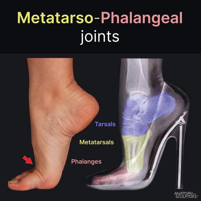
Have you ever wondered: “How do they walk in heels so high?” Standing on your toes requires plantar flexion. Tarsal and metatarsal bones move downward while phalanges stay roughly in the same position. This is possible because of the metatarsophalangeal joints. #anatomy4sculptors
The under-eye area between the lower eyelid and the inferior infraorbital margin is called the tear bag. Most anatomy sources consider it a part of the lower eyelid, but for artists, it’s essential to make the distinction and understand the separate forms. #anatomy4sculptors
Instead of being a six-pack, ABS are actually an eight-pack – four pairs of muscles, identical on each side of the linea alba. They are sometimes mistakenly depicted as rectangles. In reality, as seen in the picture, their form is much curvier. #anatomy4sculptors
Walking is one of the most common things we do. Lifting the heel involves a type of motion called plantar flexion. It changes the surface form of the ankle due to the flexion of the peroneus longus and peroneus brevis muscles. #anatomy4sculptors
The bony bump over the side of the hip is the greater trochanter – one of the most prominent bony landmarks of the femur. The side-by-sides illustrate its location on the skeleton and its position among the forms of the surface anatomy (side and hind views). #anatomy4sculptors
This illustration shows a color-coded skeleton and surface anatomy of the bust with the bony landmarks in a corresponding color code. These include the sternum, clavicle, acromion of the scapula, and hyoid bone. And also the 1st and the 2nd pair of ribs. #anatomy4sculptors
When the arm is abducted, the scapula rotates laterally in relation to the thorax. The medial border of scapula indicates the degree of this rotation. The movement is created by the Scapular muscles, Latissimus dorsi, Teres major, and Serratus anterior. #anatomy4sculptors


