histologicalのTwitterイラスト検索結果。 5 件

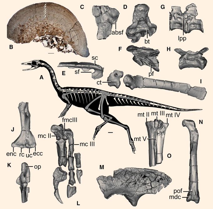
is the skeletal anatomy of Bannykus wulatensis: (A) skeletal silhouette showing preserved bones (in gray); (B) histological thin-section of the fibula; (C) left frontal in dorsal view; (D) basioccipital in ventral view; (E) left surangular in lateral view; (F-H) vertebrae in left


These teeth aren't histologically like mammals', it's basically just chitin, much like snail shells.
I present to you this cursed Wikipedia diagram how a snail eats.
I don't even know how to start describing this as alt-text, but I promise you it's probably for the best🐌🧵3/4


T cells are trained in thymus. Take a look at the histological look of their military college with @smlungpathguy & @Path_Matt https://t.co/fY5lVoG5pp

New specimens of the suchian archosaur Gracilisuchus from the Triassic of Argentina- nice forelimb material and histological analysis by #DAWNDINOS collaborator Lecuona et al.: https://t.co/mpMkijPDeK


Airheads: Comprehensive description and comparison of the complete head #pneumatic systems of the Japanese quail & the zebra finch using soft tissue enhanced micro-CT with 3D reconstructions & #histological sections. See it in the July issue!
https://t.co/If5Pk9myus