microctのTwitterイラスト検索結果。 12 件

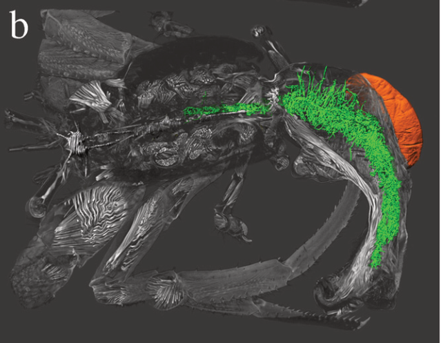
This is a microCT scan of an infected hermit crab showing the external blob part (orange) and the internal root-like barnacle tissue (green). Other parasitic barnacles have different strategies...
image from: https://t.co/USH8TEadcU


This thin but dense outer layer is tessellated prismatic cartilage, composed of small blocks (tesserae) of calcified cartilage. This calcification gives the cartilage much of its strength and makes it more radiodense (dense to X-rays). (2/4)
#sharks #CTscanning #microCT #anatomy


Slow moving progress on this, but I finally finished segmenting all of the external features in this Aeolus mellillus #microCT scan! Starting on the muscle today 💪


Day 26 Favorite Color
Same thing like the day 24 challange but this time use only ONE favorite color and draw it in monocromatic..
#Microctober2021
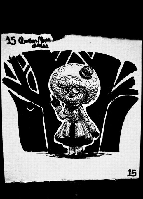
Day 15 Queen/King Dress
Doesnt look like queen dress but all i did is searching on google Queen dress and bunch clothings appears to choose..thats the challange
(Also,this character (Trilia) is a queen)
#Microctober2021


Curious what's between the broadly overlapping / #telescoping bones of dolphin skulls? For some cool histology & microCT check out my #SICB2021 talk! Live Discussion TOMORROW @ 10:30am EST / 7:30am PST, Spines & Sutures. Collab. btwn Roth lab, @MatthewJHilton, & UNCW #VABLAB


Finding that segmenting microCT data of the early dinosauriform Marasuchus/Lagosuchus for #DAWNDINOS is just what I need right now. Very relaxing, and my skills have improved a lot, so it's satisfying too. Left hindlimb here; pieces done, now to put it back together in 3D.

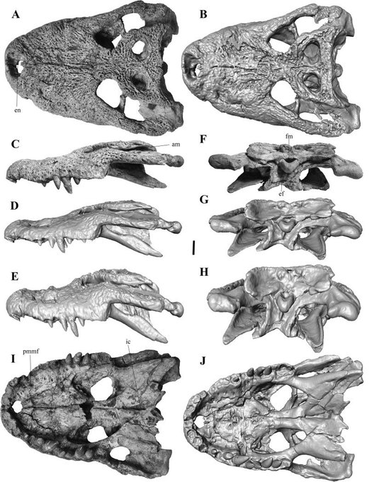

Great paper (https://t.co/maF9IARGHk) on the inner skull cavities of #eusuchian #crocodyliforms from the upper #Cretaceous 🐊Find one of the extant comparative specimens here: https://t.co/LB43OHCKrp #FossilFriday #crocodiles #skull #microCT @inarvaezp @PalaeoStephan @frco_ortega


2/5
2014 – it’s moving! Interactive animated Trigonopterus #weevil; scan duration ca. 50 min; manual segmentation of tomographic volume with Amira; animation and rendering with Deep Exploration. https://t.co/Z9m3CoMXCv
#microCT #3D #animation #beetle


Here is a (transparent) rotational gif of the skeleton of the holotype of Rhombophryne vaventy, the first species I ever described, with Zoosphaerium sp. in its gut. I just added this to @WikiCommons as CC-BY-SA 4.0 and to https://t.co/Cohb0Ivo68
#microCT #µCT #osteology #Science


MicroCT viz of a donated Carpet python #SerpentesSaturday. More to come!

