ventricleのTwitterイラスト検索結果。 42 件
If you have not heard of the
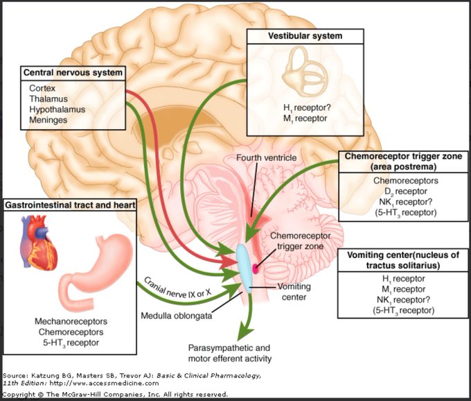
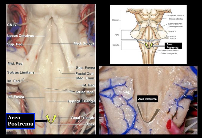
AREA POSTREMA, you'll never forget it. It's located on the dorsal surface of the medulla at the floor of the 4th Ventricle, and it detects emetic toxins in the blood & CSF and induces vomiting.
From the length of the needle and the degree of oscillation, Callender inferred that it had pierced the heart just above the apex - near the tip of the left ventricle.
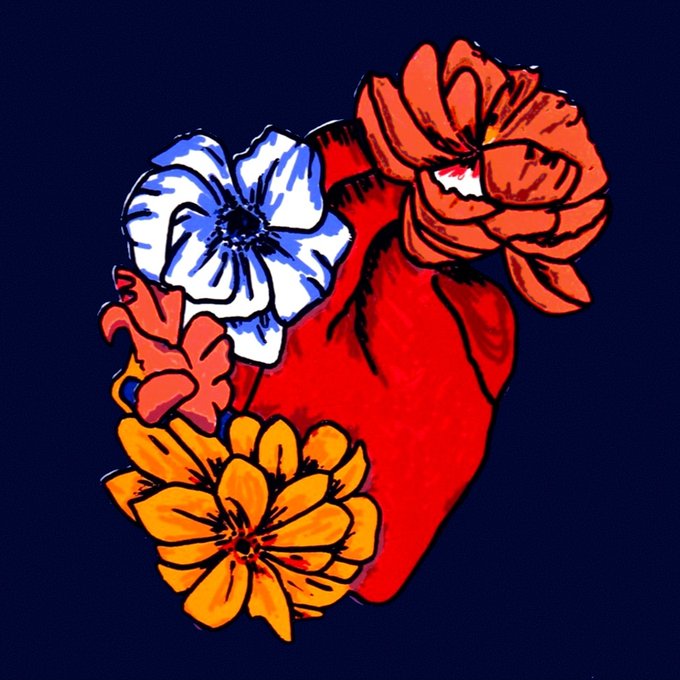
It's not always about #hearts & #flowers... but sometimes it is! This flowering pair of ventricles is "Bloom" by Katy Herbert.
This #LimitedEdition #screenprint has a beautiful subtle #iridescent #ink background, with #fluoro details. Pumping away at https://t.co/W1hypOtGCy
If you have not heard of the AREA POSTREMA, you'll never forget it. It's located on the dorsal surface of the medulla at the floor of the 4th Ventricle, and it detects emetic toxins in the blood & CSF and induces vomiting.
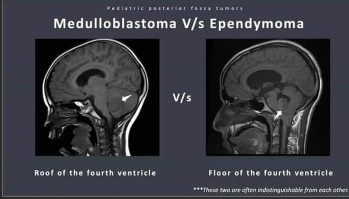
#Neurosurgery students: before resecting p-fossa tumors on peds service, know all presentations, Ddx, differentiating these lesions on CT & MRI, surgical approaches, 4th ventricle anatomy, adjuvant treatments, genetic abnormalities, postop issues mutism/hydro/other, & prognosis.
@BitmanTW The partner pieces in the Ventricles collection are now live! “Little Do We Know” and “Kezazah or the Unbearable Weight of Expectation.”
https://t.co/JsF5jG1nAk
⬇️NEW DROP⬇️
The third drop of the VENTRICLES collection is live!
"Kezazah or The Unbearable Weight of Expectation"
A lifetime of struggle under the external pressure of assumptions...we can all relate.
3/3 editions, .12ETH
#nftartist #nftart
https://t.co/YnIWY5xAd4
Thank you, @JAC_INK for picking up a copy of “Little Do We Know” from the Ventricles collection. Ecstatic to welcome you to the family!
1/3 editions left!
#nftcollector #NFTcollection #NFTart
https://t.co/iRLXaerQKQ
Things which bother me a lot in media, art, films, everything.
People pointing to the left side “this is where your heart is”
The human heart is in the center of your chest. The left ventricle is larger than the right so it leans left in that side but the heart is in the middle.
Cross-sectional view of the uterus and bladder in situ (left) and an illustration of a colloid cyst in the third ventricle of the brain (right). By one of our in-house artists. © Evan Oto/Science Source https://t.co/CZgbQnVb5n #medicalillustration #stockillustration #medical
Work in progress depicting the transcallosal transchoroidal approach to cavernous malformations of the periaqueductal gray matter in the third ventricle. The choroid plexus is my new favorite thing to draw in Photoshop 🍇 #medtwitter #neurotwitter #neuroscience
I just can't get over the beauty of DTI scans... 🤩
Check out this 3D model that includes fibre tracts, ventricles, and deep forebrain structures:
https://t.co/tMXnFRKZYm
@HiveUBCMedicine
Our illustration of the temporal horn's coronal cut; showing tapedial fibers, collateral eminence, and sulcus for Dr. Orhun M. Cevik's presentation about selective amygdalohippocampectomy.
#medicalillustration #sciart #neuro #medicalart #drawingdoctors #neuroanatomy #ventricle
Blame Double (Skullgirls) for making me getting interest drawing body horror.
Random character, named her Aneurysm. She's all ventricles and arteries.
I sometimes like making themical/terminological characters...
#originalart #veins #randomcharacter #bodyhorror
A planned echocardiogram was abandoned when he started to sweat violently, his blood pressure dropped and his jugulars became distended. Cardiac tamponade, you cry! And so he was whisked into surgery, where a large excoriation was found in the wall of the left ventricle.
(2) Meninges: meningitis, ependymitis. (3)Extra-axial which includes extradural or subdural empyema (4) ventricles and includes ventriculitis. #BBLive
Throwback: Beautiful traditional drawing from the @BarrowNeuro archive, by Steven J. Harrison, PhD, circa July 1989. It depicts a suprasellar tumor invading the third ventricle. Steve used airbrush, gouache, and color pencil to bring this piece to life. #traditionalart
Seen here are the operative viewing angles (green) and blind spots (red) encountered during a transforaminal transvenous transchoroidal approach, which exposes the third ventricle by enlarging the foramen of Monroe via transection of the septal vein.
https://t.co/Rn1NHc1Ezr


