PDBのTwitterイラスト検索結果。 222 件中 7ページ目
Remdesivir, Sofosbuvir
NS5B RdRp with Sofosbuvir (4WTG.PDB)
SARS RdRp homology model (1SXF.PDB)
核酸の塩基をミミックしている部分に違いがあるな!
RdRp 同士、フォールディングは近いが活性部位には個性がある。
Ectodysplasin-A is recognised by EDAR, @ensembl's #geneoftheweek, and involved in signalling cell death. There are 2 structures in the PDB, representing 2 isoforms - binding to either EDAR or EDA2R. View the structures at #PDBeKB: https://t.co/JIS2po2O5u https://t.co/dgU0HbmMGr
#Drosophila dArc proteins can built retrovirus-like capsids! dArc 1 and 2 are used for #synaptic plasticity in Drosophila. Published 2020 @NatureNeuro. PDB: 6TAP
#scicomm #sciart #b3d #retrovirus #Gag_gene
PDB images often appear in textbooks, flyers and posters. Bonnie Hall (@GrandViewUniv) describes how initial interest in these images can be leveraged to engage students in a variety of chemistry classes https://t.co/MymAigk6Ms
MERRY (PDB ID: 2iuu, residues 535-539) CHRISTMAS (PDB ID: 2m0m, residues 34-42, required 3 mutations) @mrc_hgu @MRC_IGMM @buildmodels
This week's release sees 240 new PDB entries released into the wild! One of these is the structure of the major strawberry allergen, published in J. Agric. Food Chem. (@ACSPublications). Find all the new entries at https://t.co/54E03LeH5m
Explore PDB-101 resources on Nobel Prize awards that recognized achievements made in molecular biology, structural biology, and related research https://t.co/ACbcfVnSh4
Happy birthday to Thomas Cech, who won the Nobel prize for his discovery of catalytic RNA. He has 17 entries in the PDB, including this structure of the Tetrahymena ribozyme. View the structure using Mol* at https://t.co/V8gRdHZYle
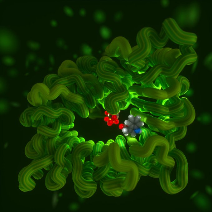
New in-browser molecular viewer Mol* from @PDBeurope, RCSB PDB @buildmodels , and @CEITEC_Brno! I took it for a quick spin to visualize a recent release, PDB 6T8G. Love that it includes AO! @AMIdotorg #sciart https://t.co/fyiND6iiMj
Enzyme adds iodine to diverse substrates https://t.co/N0B2wj3vMU via @cenmag Explore in 3D at RCSB PDB: https://t.co/Ol9U7e4rPx
There are 235 new entries in the #PDB this week, including this giant EM structure of S. aureus bacteriophage P68 from @PlevkaLab - published @ScienceAdvances. Find all this week's entries at https://t.co/54E03LeH5m
Pranlukast, a drug for treating asthma arrives in the PDB today, bound to it target the cysteinyl leukotriene receptor.
Solved @esrfsynchrotron and published in @ScienceAdvances, it's one of the new structures today at https://t.co/54E03LeH5m
David Goodsellさん@dsgoodsellが使っている立体構造イラストソフトがwebベースで使えるようになったんですね!
Thermococcus litoralis 4-α-glucanotransferase (pdb: 1K1Y)
大学院生の時に構造決定した思い出のタンパク質をGoodsellさん風に。 https://t.co/XchxV7VdEU
Constantin Fahlberg was granted a patent #OTD in 1885 for the use of artificial sweetener saccharin, which is found bound to 3 different proteins in the PDB
https://t.co/Fy0qMiJUmJ
The little molecule is the most abundant auxin - a plant hormone. Here it is inside the plant, inside the F-box protein TIR1 ubiquitin ligase
suggested for my #molecularportrait s by @DKampjut, tnx🙂
Struct ref: doi:10.2210/pdb2P1Q/pdb
👇pls suggest what to see next
#sciart
Yep. David S. Goodsell made "Illustrate: Non-photorealistic Biomolecular Illustration" so you can illustrate any PDB id.
https://t.co/nQOQovoEse
This one I made of the cryptochrome 4I6E.
Great tool from David Goodsell for the creation of protein structure illustrations in the typical style of the "PDB Molecule of the Month". Here is an example using the methanol dehydrogenase from M. extorquens. https://t.co/woZC79oMUq
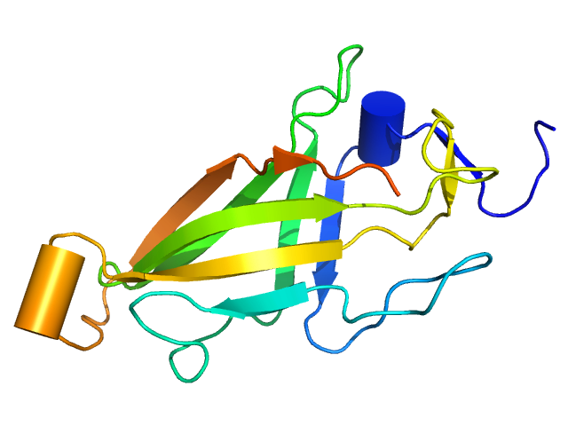
And its a new fold! The “SARAF-fold" is novel 10-stranded β-sandwich fold which domain swaps to dimerise.
#PDBeFold confirms there are no similar structures in the PDB archive


