cranialのTwitterイラスト検索結果。 249 件中 10ページ目


More evidence for terrestriality in baurusuchids. Great to see notosuchian endocast/endocranial morphology being more explored!
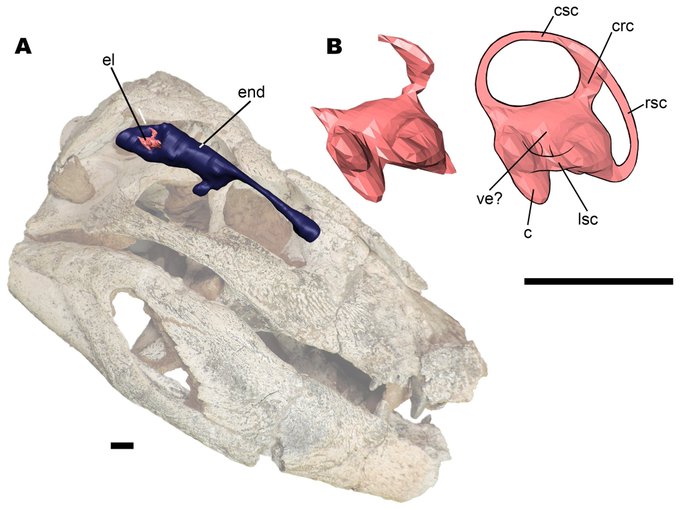
Fonseca et al. (2020) Morphology of the endocranial cavities of Campinasuchus dinizi from the Upper Cretaceous of Brazil https://t.co/8u49zcU1ep


A coronal view of the surgical trajectory for the interhemispheric corridor. Important structures include: 1) cingulum, 2) corpus callosum & 3) fornices. Take care to preserve the cerebrovascular structures at each dissection level.
#nsgy #complexcranial
https://t.co/9LBUa8OSy7
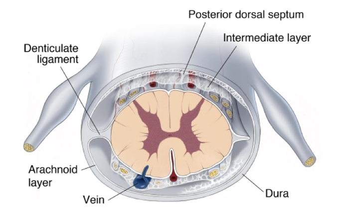
The arachnoid layer of the #spinalcord is attached to the dura. The pia is firmly attached to the dura by 21 pairs of extensions called denticulate ligaments.The denticulate ligaments promote spinal cord stability by limiting cranial-caudal movements.
https://t.co/tBbewYe6wY


#Talarurus portrait. Commissioned by the authors of a new paper describing the cranial anatomy of this #ankylosaur.
https://t.co/AtMbEE0H7a

Guanlong showed that every lineage with giants had small beginnings. Also the cranial crest of Guanlong is intriguing, and may have been used for display. Artwork by @BrianEngh_Art


Cranial vault thickness variation and inner structural organization in the StW 578 hominin cranium from Jacovec Cavern, South Africa. Source: Beaudet et al (2018)


American neurosurgeon Peter Jannetta (1932-2016) was dissecting cranial nerves in the lab when he noticed a blood vessel pressing on a nerve. This led to a microvascular decompression operation for the treatment of trigeminal neuralgia and other vascular compression syndromes!


Artists are a curious lot, heads filled with mysterious images wanting to manifest in a visual sensibility.
Imagine that.
It is amazing to consider some mental impulse traveling from the cranial cavern, through the arm and out the fingers. What an incredible process it is.
— MAFO


#MemoriaDeLaExtinción 279. Caviramus schesaplanensis. Un pterosaurio ranforrincoideo de hace 205 Ma. Tenía una mordida potente, cresta cranial pronunciada y unas alas largas y estrechas. Parecía estar bien adaptado a una vida en el suelo. (©Mark Witton)

#ImageChallenge: partially thrombotic & calcifie L ICA bifurcation aneurysm!
Patient received high-flow extracranial ICA-M2 bypass & Hunterian ligation of the ICA distal to the anterior choroidal artery.
https://t.co/MtwPhB4Lf6
#bypass #nsgy #complexcranial #cerebrovascular


Artists are a curious lot, heads filled with mysterious images wanting to manifest in a visual sensibility.
Imagine that.
It is amazing to consider that mental impulse traveling from the cranial cavern, through the arm and out the fingers. What an incredible process it is.
— MAFO

Neurovascular conflict for cranial nerve compression syndromes can include arteries and veins. Which artery is the most common culprit in trigeminal neuralgia? What about in hemifacial spasm?
#trigeminalneuralgia #MVD
https://t.co/dkhLkZZdoE

#GrandRounds: "Supracerebellar Approach: Expanding the Operative Corridor" with Dr. Aaron Cohen-Gadol
#complexcranial #nsgy
https://t.co/sL3KM4bAXh


(hopefully) got all the overlapping cranial material, although still debating on whether to include that one nasal fragment
postcrania is going to be a pain...
#spinosaurus, #sigilmassasaurus, and anything inbetween

A quite complete pelvis from 0.9–1.4 mya: BSN49/P27 from Gona, Ethiopia, attributed to Homo erectus/ergaster. A nice exception in the poor post cranial fossil record of Early Pleistocene Homo... Source: Simpson S.W. (2015) https://t.co/kQCo7DYJDU
Blue arrow demonstrates the supralateral cerebellar approach for microvascular decompression for trigeminal neuralgia. Green arrow demonstrates the infralateral cerebellar or infrafloccular approach for hemifacial spasm.
#nsgy #complexcranial @TheJNS
https://t.co/IeKulKlHtr

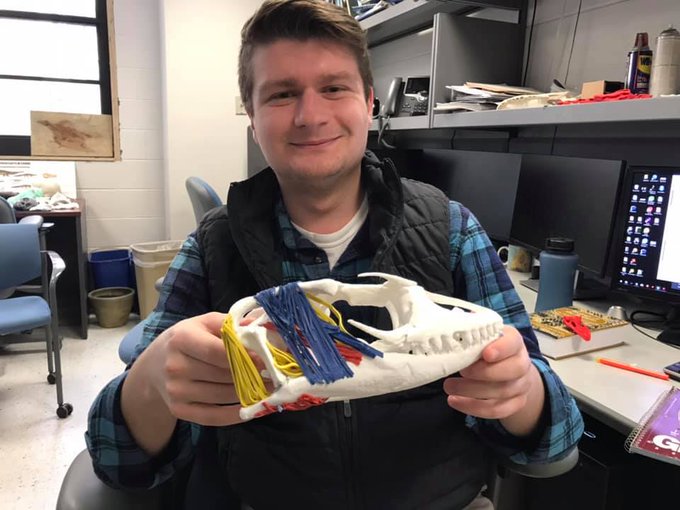
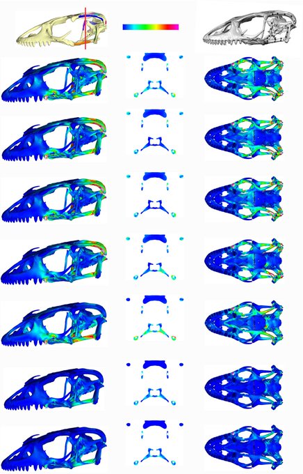
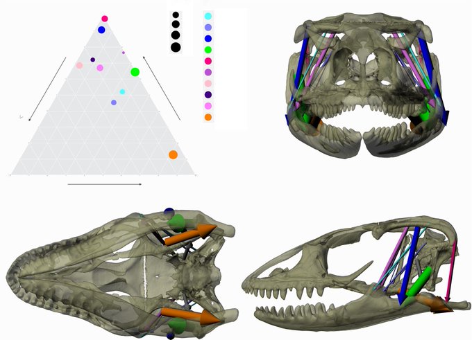
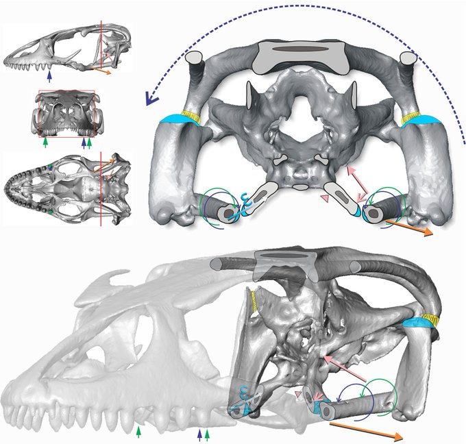
Congrats to Alec Wilken for seeing this monster of a paper through to @J_Exp_Biol. How do joints and muscles mediate cranial kinesis and torsion in the skull of a varanid lizard?https://t.co/uV3gdDm0Yf @mizzouanatomy @mizzou @NSF_BIO @MizzouNews @MizzouBiology @ugradresearchMU

Endocranial shape Neandertals v. Modern Humans: A=Nea, La Chapelle-aux-Saints, elongated. B=MH, globular. Arrows=enlarged posterior cranial fossa (housing the cerebellum) & bulging of parietal bones in MHs compared to Nea. Source: Gunz et al (2019) https://t.co/TrdnMd91he


THE CRANIAL ALPHA
Personnel image from a script I have been writing. This is long term project as it would be damn expensive to make.