NsgyのTwitterイラスト検索結果。 34 件中 2ページ目

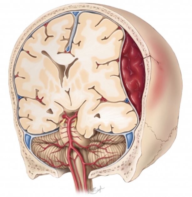
A large epidural #hematoma (#EDH) causing mass effect is demonstrated. Which types of #herniation could be caused by a large, expanding EDH?
#nsgy #trauma
https://t.co/l2xTG8so43
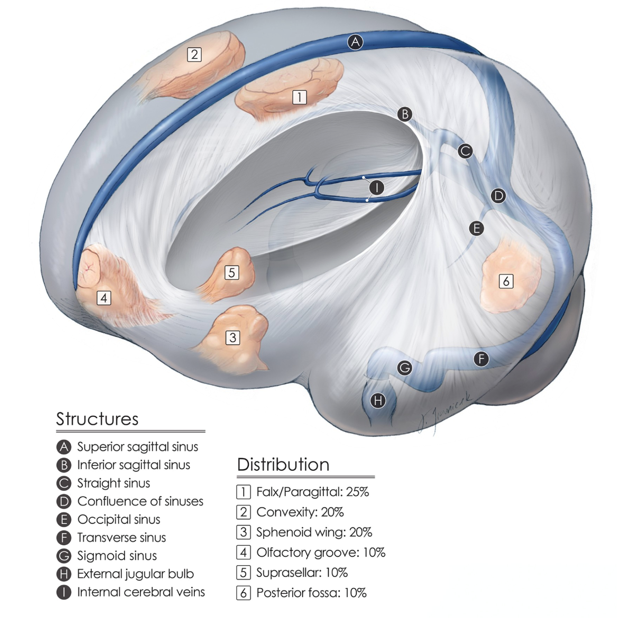
Here, we see the relative distribution of the most common intracranial meningiomas. Resection of these benign tumors is associated with good patient outcomes and therefore is satisfying for neurosurgeons.
#nsgy #tumors #meningioma
https://t.co/Ldg5aBh08O

A coronal view of the surgical trajectory for the interhemispheric corridor. Important structures include: 1) cingulum, 2) corpus callosum & 3) fornices. Take care to preserve the cerebrovascular structures at each dissection level.
#nsgy #complexcranial
https://t.co/9LBUa8OSy7

During the telovelar approach, the foramen magnum is unroofed & burr holes are placed on either side of the midline keel.The tonsils, vermis, & medial cerebellar hemispheres are exposed. Patent occipital sinuses can cause brisk bleeding.
#nsgy
https://t.co/Gcl58A4kWM

When debulking a fourth ventricle tumor, it is critical to preserve the floor of the fourth ventricle, even if that requires leaving behind a carpet of invading tumor. Can you name some common fourth ventricular tumors?
#nsgy #tumors
https://t.co/XGvc0ts2Sf


Merry Christmas and Happy New Year from all of us @QSReview!
We’re looking forward to seeing many of your at #QSNS2020!
@The_SBNS @EANSonline @CNS_Update @WFNSHQ @AANSNeuro
#neurosurgery #nsgy

Injury to the anterior 1/3 of the superior sagittal sinus (SSS) can be repaired by tying off the proximal and distal ends of the tear with 2-0 silk sutures.
#sagitalsinus #repair #nsgy
https://t.co/69Xj4lV2Ae

#ImageChallenge: partially thrombotic & calcifie L ICA bifurcation aneurysm!
Patient received high-flow extracranial ICA-M2 bypass & Hunterian ligation of the ICA distal to the anterior choroidal artery.
https://t.co/MtwPhB4Lf6
#bypass #nsgy #complexcranial #cerebrovascular

#GrandRounds: "Supracerebellar Approach: Expanding the Operative Corridor" with Dr. Aaron Cohen-Gadol
#complexcranial #nsgy
https://t.co/sL3KM4bAXh

The #imagechallenge from yesterday was a spinal cavernous malformation (CM)!
The image below is depicting techniques for their removal with full explanation found at the link below:
https://t.co/Bqk7wUUDnO
#cavmal #nsgy #spine #surgery

The peri-insular hemispherotomy effectively treats patients with unilateral epileptogenic hemisphere dysfunction while limiting potential complications. This illustration shows the exposure of the frontal horn of the lateral ventricle.
#epilepsy #nsgy
https://t.co/YlXCWBjAXD
Blue arrow demonstrates the supralateral cerebellar approach for microvascular decompression for trigeminal neuralgia. Green arrow demonstrates the infralateral cerebellar or infrafloccular approach for hemifacial spasm.
#nsgy #complexcranial @TheJNS
https://t.co/IeKulKlHtr

Demonstration of the removal of a hypothalamic hamartoma (blue arrows define the border of the lesion). It is imperative that the hamartoma-hypothalamic interface is left untouched. What is the hallmark clinical sign associated with these lesions? #nsgy
https://t.co/wT2dl3fI8q

Illustration depicting the typical appearance of a cavernous malformation. #brain #study #neuro #learning #nsgy #medstudent
https://t.co/g53roOPqtE