micrographのTwitterイラスト検索結果。 95 件中 4ページ目


NIKON SMALL WORLD PHOTOMICROGRAPHY 2020
1st Daniel Castranova, Dr Brant Weinstein & Bakary Samasa: Juvenile zebrafish
2nd Daniel Knop: Embryonic development of a clownfish on days 1, 3, 5 and 9
3rd Dr Igor Siwanowicz: The tongue of a freshwater snail
https://t.co/0QOgHaDitX


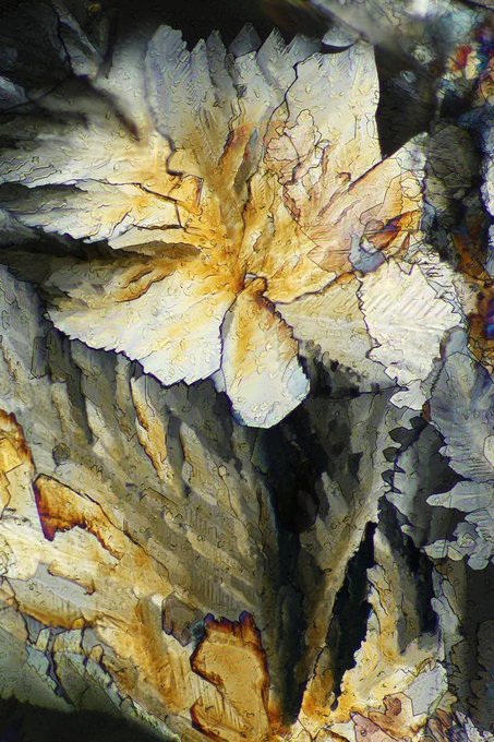


#DidYouKnow chocolate looks as blissful as it tastes? 🤩🔬
Lee Hendrickson's #photomicrography of crystals include intricate structures from wine, chocolate, caffeine, citric acid, to acetaminophen that transform into vivid colors and mesmerizing patterns. https://t.co/iLREcztTKC


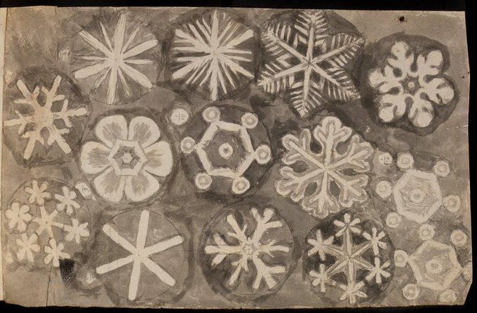
In 1665 Robert Hooke published his most famous work Micrographia (”Small Drawings”). In it he included his studies and illustrations of the crystal structure of snowflakes and first used the word cell to name the microscopic honeycomb cavities in cork.5/


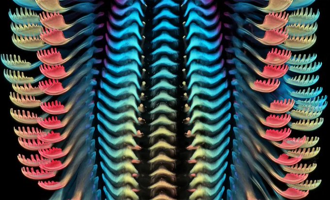
Tortoise Beetle, SEM
Color enhanced scanning electron micrograph (SEM) of the underside of a tortoise beetle (family Cassidini)
© Steve Gschmeissner/Science Source
Science Source ID# SS2310805
#beetle #tortoisebeetle
https://t.co/2IPDRc4ENX


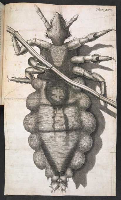
@17thCenturyLady @jdmccafferty @RestorationHat @DrRJWarren @saralread @tomb1649 @kristianjmartin @quintonjournals @cheapsellotape Robert Hooke, FRS (1635-1703), co-inventor of the watch balance spring, architect, but remembered best for his Micrographia (1665) - you'll have seen his Flea.
Worked with Boyle, Newton et al. #StuartsSaturday #KeepItStuart

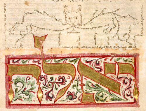

Decorated initial-word panel from the opening of Shemot (Exodus) with micrography from masoretic notes for
#MicrographyMonday
Add 19776; the 'Coburg Pentateuch'; Germany, Central (Coburg); 1390-1396; f.28v @BL_HebrewMSS
@BLAsia_Africa


At the #SICB2019 #Buchsbaum Photomicrography Contest, our new micro-CT category was full of popular submissions, including these 3D scans of leather star (Dermasterias imbricata) and slime star (Pteraster tesselatus) endoskeletons by @PycnoPod and @CMDonatelli! #Scanallstars

Polarizing light micrograph of Vitamin E crystals.
#VitaminE is a fat-soluble nutrient found in many foods. In the body, it acts as an antioxidant, helping to protect cells from the damage caused by free radicals. #FunFactFriday #FridayThoughts #science #medical #images #photos


It was #OnThisDay in 1664, the great British natural philosopher Robert Hooke FRS, first showed his copy of the hugely influential Micrographia to the Royal Society. Find prints from this seminal work in our Print Shop: https://t.co/OkGAr01G5r


Fluorescence confocal micrograph of human cerebral organoids labeled with markers of neuroepithelial cells, neuronal progenitors, and cortical neurons, by Laura Pellegrini @MRC_LMB from this @CellPressNews picture show: https://t.co/fgnYEd9ITA

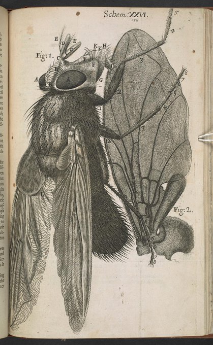

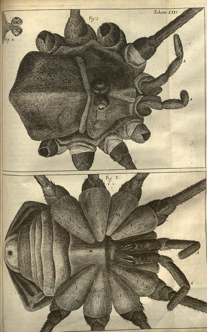
Micrographia, or some physiological descriptions of minute bodies made by magnifying glasses, with observations and inquiries thereupon.
https://t.co/dKh5IqgykR


Wow! This is a micrograph of a butterfly's tongue magnified 720 times. Looks like #embroidery


Pollen micrograph by Andrew Paul Leonard https://t.co/yjaebLoqPn #sciart


Microscopes meet Indigenous art in Stories & Structures – New Connections, a stunning new exhibition by @GoInnerSpace: https://t.co/SnwSQOfbPM #SciArt #scicomm #paintings #Indigenous #art #science #micrography


This photomicrograph shows the fine brushwork Jacob van Walscapelle used to paint a ladybird. Each leg is only 2mm. #BSW18 @ScienceWeekUK


Robert Hooke, English natural philosopher, architect and polymath (author of 'Micrographia') died #OnThisDay in 1703.


This thread is a series of images in the #history of #scicomm documenting the shift in 17th century Europe from divine causation to empiricism (via observation, experiment, reasoning, & formation/testing of hypotheses & theories).
[Micrographia, 1665 https://t.co/1202Hyolle ]


It's cold out there today! We've got Hooke on our minds at the #T3D2019 labs, here's a few #T3D2019_PoftheD from his Micrographia (1665) and his 1662 article in @royalsociety Philosophical Transactions

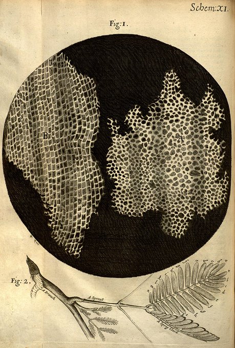
Robert Hooke was born on 28 July 1635 N.S. His Micrographia includes the 1st use of "cell" as a biological term https://t.co/9YXRp9beYw