AVMsのTwitterイラスト検索結果。 8 件


September is #Aneurysm AwarenessMonth. This sequence of illustrations was created for The article, “The future of open vascular neurosurgery: perspectives on cavernous malformations, AVMs, and bypasses for complex aneurysms” The article is free access: https://t.co/M3vKfDghtm


Here @AaronCohenGadol describes the resection of AVMs within eloquent cortices and more specifically the angular gyrus or language cortex #Nsgy #AVM https://t.co/hNV5m9qJiP
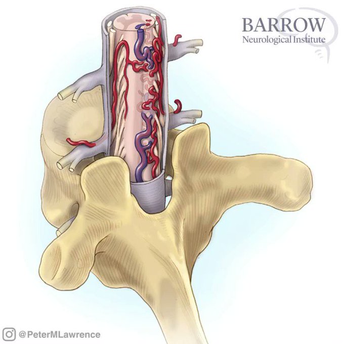
#Throwback circa 2002. Illustrations from @BarrowNeuro medical illustrator and manager of Neuroscience Publications, Mark Schornak. Depicts spinal AVM classification of compact intramedullary spinal AVMs (thoracic vertebra, posterior view)@BipinChaurasia_ @neurocirurgiabr


Watch as Dr. Cohen removes an arteriovenous malformation within eloquent cortices using the intranidal resection technique. This technique allows for maximal protection of vital structures around the nidus.
https://t.co/JZhxEV9GO2
#AVMs #cerebrovascular #nsgy

Temporal horn AVMs are technically challenging to tackle because the nidus is covering the feeding vessels emerging from the anterior choroidal artery within the choroidal fissure. #nsgy #neurosurgery #cerebrovascular
Learn more here:
https://t.co/cn2k9dzhwt

For ventricular body AVMs that extend lateral to the midline, a contralateral transcallosal route is favored to minimize ipsilateral hemispheric retraction. #nsgy #neurosurgery #cerebrovascular
Learn more here:
https://t.co/cn2k9dzhwt

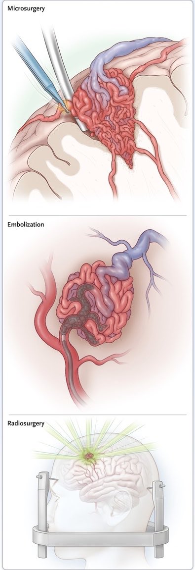
“AVMs: What, how, why and when?”
There’s definitely no “who” in that title because of course it’s @PatrickGrover4 (@QSNeurosurgery) and it’s only a week away!
Wednesday 29th Jan, 7pm, @RCSnews.
Get excited! #neurosurgery
(Pic courtesy of @NEJM - https://t.co/HaPVBgBitY)

Illustration depicting the en bloc removal of a hemangioblastoma. The deep white matter feeders often present in AVMs are not present in hemangioblastoma resections, which aids in greatly simplifying the procedure. #braintumor #neuro #brain #neurosurgery
https://t.co/mKFEinhyPU