
Neurosurgical Atlasさんのイラストまとめ
@neurosurgatlasFollow @neurosurgatlasさんをフォローする
フォロー数:818 フォロワー数:26380


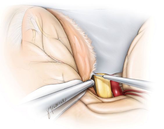
During resection of cavernous sinus meningiomas, first remove the components of the tumor affecting the optic nerve to protect the nerve during tumor manipulation & traction. Note aggressive devascularization of the tumor & its thorough internal debulking.
https://t.co/jHZE5rGYn1

During transcranial resection of tuberculum sella meningiomas, it's crucial to preserve the small branching vessels originating from the superior hypophyseal artery complex & the medial aspect of the internal carotid artery supplying the optic nerve/chiasm
https://t.co/vyQXmbNxZ3
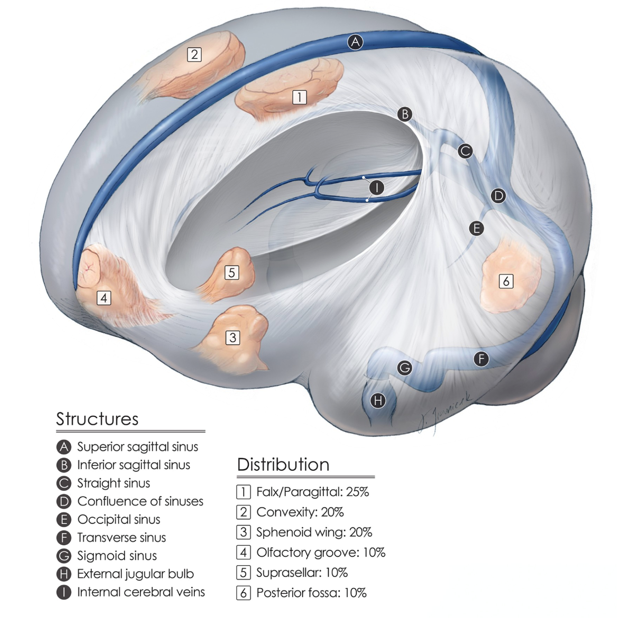
Here, we see the relative distribution of the most common intracranial meningiomas. Resection of these benign tumors is associated with good patient outcomes and therefore is satisfying for neurosurgeons.
#nsgy #tumors #meningioma
https://t.co/Ldg5aBh08O
While resecting olfactory groove meningiomas, I dissect the arachnoid plane between the ipsilateral optic nerve/chiasm/carotid artery & the posterior capsule of the tumor. Early ID of these structures improves the efficiency & safety of the operation.
https://t.co/Ldg5aBh08O

A transcondylar approach w/ C1 hemilaminectomy for approaching a foramen magnum #meningioma. The relationship between the vertebral artery and tumor is important. Most such tumors can be resected via a lateral suboccipital approach without a condylectomy.
https://t.co/TzVD3mfYlj
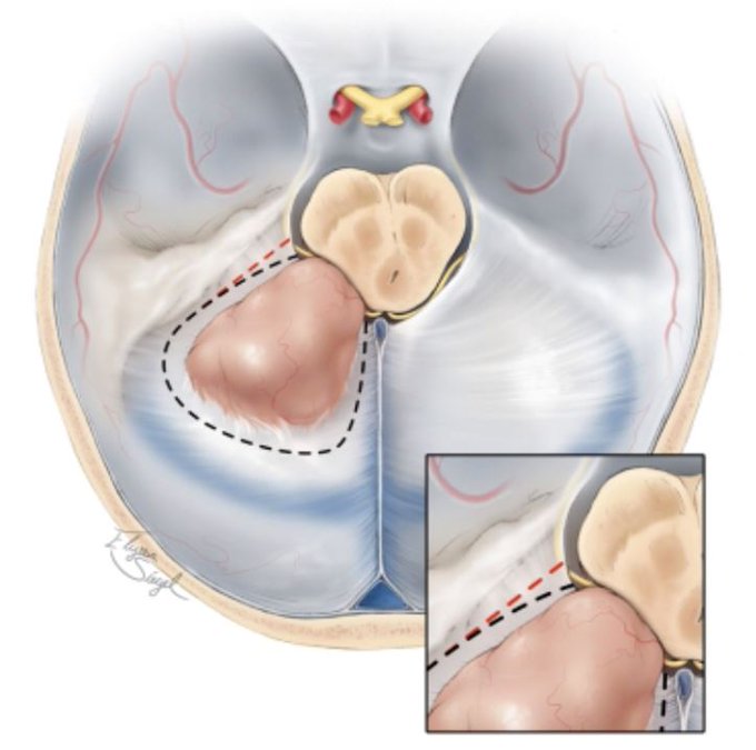
During tentorial transection for a petrotentorial #meningioma, it's important to protect the trochlear nerve as it travels along the lateral edge of the incisura. The black dotted line represents an incision that allows us to avoid an injury to the nerve.
https://t.co/73xKJbX8vV

Olfactory groove meningiomas can be resected via a bifrontal craniotomy. I prefer a pterional craniotomy, no matter how large the olfactory groove meningioma. This approach is less invasive & allows early identification of the optic nerve & carotid artery
https://t.co/Ldg5aBh08O

A coronal view of the surgical trajectory for the interhemispheric corridor. Important structures include: 1) cingulum, 2) corpus callosum & 3) fornices. Take care to preserve the cerebrovascular structures at each dissection level.
#nsgy #complexcranial
https://t.co/9LBUa8OSy7
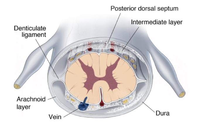
The arachnoid layer of the #spinalcord is attached to the dura. The pia is firmly attached to the dura by 21 pairs of extensions called denticulate ligaments.The denticulate ligaments promote spinal cord stability by limiting cranial-caudal movements.
https://t.co/tBbewYe6wY

During the telovelar approach, the foramen magnum is unroofed & burr holes are placed on either side of the midline keel.The tonsils, vermis, & medial cerebellar hemispheres are exposed. Patent occipital sinuses can cause brisk bleeding.
#nsgy
https://t.co/Gcl58A4kWM