
Neurosurgical Atlasさんのイラストまとめ
@neurosurgatlasFollow @neurosurgatlasさんをフォローする
フォロー数:818 フォロワー数:26380

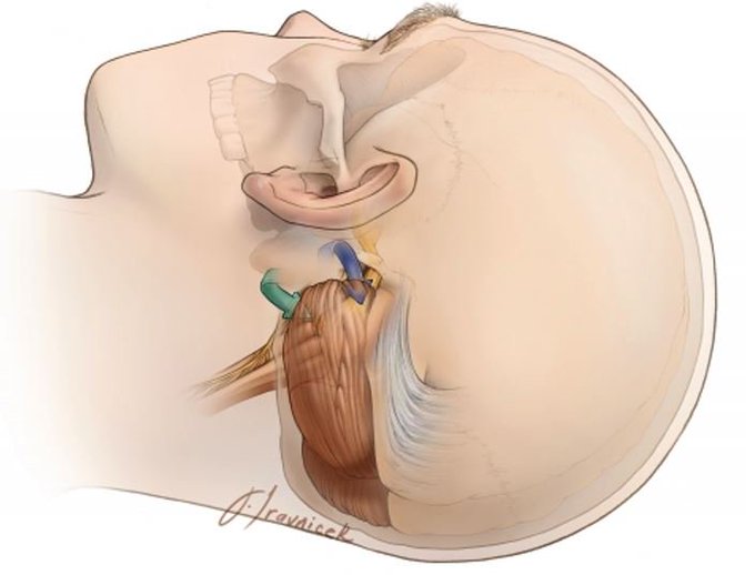
Blue arrow demonstrates the supralateral cerebellar approach for microvascular decompression for trigeminal neuralgia. Green arrow demonstrates the infralateral cerebellar or infrafloccular approach for hemifacial spasm.
#nsgy #complexcranial @TheJNS
https://t.co/IeKulKlHtr

Frontal lobectomy can be performed in certain epilepsy disorders. The picture demonstrates the oblique line for the resection, which extends from the coronal suture to the anterior skull base. Which critical structure should a neurosurgeon avoid?
https://t.co/6WQjqa75v5

Meningeal manipulation can be a source of post-operative pain for patients and bipolar coagulation should be minimized whenever possible. Here we demonstrate the nerves that innervate specific portions of the dura.
https://t.co/RZws7JxABV
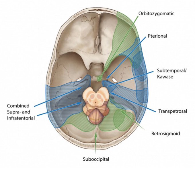
Brainstem lesions can be approached through specific craniotomies. Tell us below some lesions you’ve encountered/observed and which approach you utilized/observed!
https://t.co/12TkdsgND0
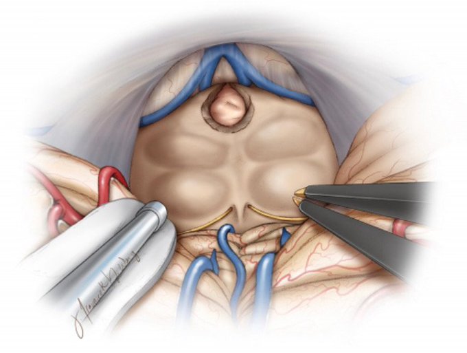
Here we examine removal of a 3rd ventricular tumor directly superior to the quadrigeminal plate. What classic syndrome could a tumor in this location cause?
https://t.co/1zAOV1Crte
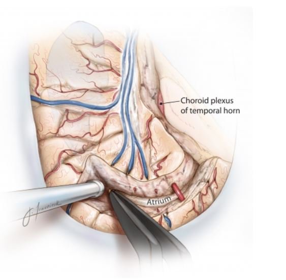
When performing a peri-insular hemispherotomy for drug-resistant epilepsy, it is important to identify the choroidal fissure because all structures located medial to the fissure belong to the diencephalon and should be protected throughout the procedure.
https://t.co/YlXCWBjAXD

Heading into an ETV this week? Check out our volume detailing the anatomy of the ventricular system. This illustration shows a superior view of the ventricles with the veins most associated with operative procedures clearly marked.
https://t.co/B26S3X0cZV

The majority of craniopharyngiomas have a large suprasellar component with cystic expansion displacing or entering the third ventricle. Shown here is the typical appearance of a retrochiasmatic craniopharyngioma.
https://t.co/fuJYioaPK2

Demonstration of the removal of a hypothalamic hamartoma (blue arrows define the border of the lesion). It is imperative that the hamartoma-hypothalamic interface is left untouched. What is the hallmark clinical sign associated with these lesions? #nsgy
https://t.co/wT2dl3fI8q

Metastatic tumors can create edema far out of proportion to their size. Anybody know the most common region for metastatic tumors to develop in the brain? #brain #neurosurgery #QandA #neuro #medicalstudent
https://t.co/56zHCKolEV