
Brainbookさんのイラストまとめ
@realbrainbookFollow @realbrainbookさんをフォローする
フォロー数:1269 フォロワー数:8596


MRI is one of the key investigations used to diagnose neurological/neurosurgical pathology. This article by @chidera_ota and illustrated @ladvic details how an MRI works and what to expect. #MRI #Nsurg #SoMe #PreMed #Medicine #neuro
https://t.co/FHVUfJUMAW

One of the key investigations carried out in neurology and neurosurgery is the lumbar puncture. This article by @Kiren_Parkash demonstrates the indications for the procedure and how it is performed! A very insightful article for patients & professionals
https://t.co/X6M8oqAUDR

35. Our patient was taken to theatre and clipping of her aneurysm was successfully performed #bbsah

23. Over time the aneurysms weaken and can rupture. When it ruptures, the blood extravasates into the subarachnoid space. This is the space between the arachnoid membrane and the pia mater. This is why we call it SAH. #bbsah art by @artibiotics

22. Aneurysms can either be saccular or fusiform in shape. Fusiform shaped aneurysms bulge out on all sides of the blood vessels. Saccular (berry) aneurysms bulge out on only one side. These are the most common type. #bbsah

17) Between the skull and the dura mater💀🧠The dura mater is the outermost layer of meninges. Meninges are the membranous covering of the brain and spinal cord. The dura is thick, tough and inextensible and lies directly underneath the bones of the skull #BBEDH

29. When CSF passes through the foramen of Luschka this results in filling of the subarachnoid space of the cisterns and the cerebral cortex.. the level at which CSF enters the subarachnoid space is called the cerebellomedullary cistern! #BBHCP

47. Disc prolapse has a variety of causes including ageing, obesity, smoking, repetitive strain, or even certain motions e.g. twisting/ turning to lift an object. CES typically results from central disc herniation. #BBCES Image: https://t.co/KOVzx1ggLB
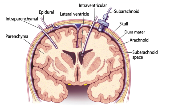
35. There are other methods to monitor ICP in different intracranial locations. These include an extraventricular drain (EVD) or intraparenchymal catheter. These are illustrated below ! #BBTBI
17. On further examination you notice her right pupil to be 7mm and non-reacting and her left pupil to be 3mm and reacting. What do you think is the cause? 🔦👀 #BBTBI