ventricleのTwitterイラスト検索結果。 46 件中 2ページ目


Seen here are the operative viewing angles (green) and blind spots (red) encountered during a transforaminal transvenous transchoroidal approach, which exposes the third ventricle by enlarging the foramen of Monroe via transection of the septal vein.
https://t.co/Rn1NHc1Ezr


Dragon's Crown_Elf by Ventricle very nice Fantasy art pixel-art! Keep it up!
#gameart #art #d #game #digitalart #gamedev #games #gaming #conceptart #illustration #fanart #drawing #videogames #gamedesign #gamer #artist #indiegame #dart #videogame #gamedevelopment #voxodyssey

The operative view through the more popular transcortical route via the left superior parietal lobule for periatrial lesions is shown with the sagittal suture parallel to the floor. Note the body of the lateral ventricle is inferior here. #nsgy
https://t.co/cn2k9dzhwt


One of my favourite anatomical terms is for the connection between the 3rd and 4th ventricles of the brain: the aqueduct of Sylvius. Named for the ancient way of moving water made famous by the Romans, and the 17th century Dutch physician Franciscus Sylvius.


Searching the outter ventricles of the Astro Shaman’s valiant Bonesteed revealed yet another creepy stowaway!
Only the owner of this piece will be able to explore the sculpture fully.
Currently only 1.5ETH on @SuperRare_co
https://t.co/w3zerQoedh


เนี่ยยยยย เห็นปั๊บคือออ Aorta, Atrium, Ventricle , Superior vena cava ลอยมาาา แต่จริงๆความติสของนางคือ จดจำทุกอย่างไว้ในใจ ทั้งคำด่าและคำชม 5555555 แต่ชอบรูปจริงๆ สวยมากๆ เป็น symbolic สุดๆ
In the telovelar approach (pictured above), removal of the C1 arch can improve visualization of the rostral half of the fourth ventricle, rivaling even that of the transvermian approach. https://t.co/FU5HnTAeSc.
#neuro #neurosurgery #surgery #brain #tumor

When debulking a fourth ventricle tumor, it is critical to preserve the floor of the fourth ventricle, even if that requires leaving behind a carpet of invading tumor. Can you name some common fourth ventricular tumors?
#nsgy #tumors
https://t.co/XGvc0ts2Sf


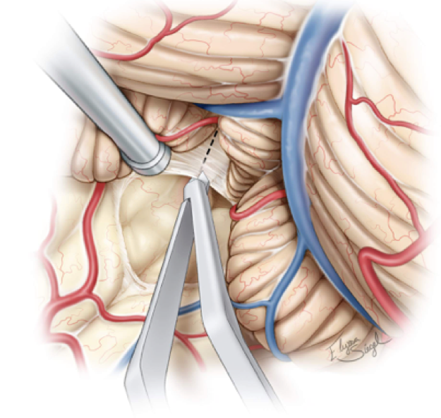
The peri-insular hemispherotomy effectively treats patients with unilateral epileptogenic hemisphere dysfunction while limiting potential complications. This illustration shows the exposure of the frontal horn of the lateral ventricle.
#epilepsy #nsgy
https://t.co/YlXCWBjAXD

Heading into an ETV this week? Check out our volume detailing the anatomy of the ventricular system. This illustration shows a superior view of the ventricles with the veins most associated with operative procedures clearly marked.
https://t.co/B26S3X0cZV

The majority of craniopharyngiomas have a large suprasellar component with cystic expansion displacing or entering the third ventricle. Shown here is the typical appearance of a retrochiasmatic craniopharyngioma.
https://t.co/fuJYioaPK2


A drawing for @Doug_Karr's novel Dwelling.
'...his heart was no longer cooperating with his ventricle chambers, which were now pumping at a ranging pace.'
See the process: https://t.co/88u5OfCr53


#JournalNC: https://t.co/yWQCXRNMSi #Machine Learning based tool to auto-segment left ventricle using #SPECT is feasible. Is it ready for prime time? #CVNuc #ArtificialIntelligence @emoryheart @MyASNC


5.Blood Flow
Trivia: The muscles of the ventricles are thicker than of the atria because they pump blood to the anterior and posterior portion of the body. Left ventricle is thicker than the right ventricle because it pumps blood to the rest of the body.


I know we all ❤️ #sharkweek, but #DYK shark hearts are different than ours? Sharks also have 4 chambers, but they're laid out in a line rather than in a square. The blood flows: body -> sinus venosus -> atrium -> ventricle -> conus arterious -> gills -> body #MorphologyMonday


Kotryna Zukauskaite
Cleveland Clinic’s Heart & Vascular Institute Milestones
Nation’s Best Heart Center - #1 since 1995
1988 Ventricle Assist Devices keep patients alive while waiting for transplants
https://t.co/moMweY5KuS
#illustration #heart #vascular #ventricleassist


Obstructive hydrocephalus due Aqueductal stenosis. Images showing an dilated third ventricle and lateral ventricels… https://t.co/cFgkT3CVhj