mskradのTwitterイラスト検索結果。 18 件
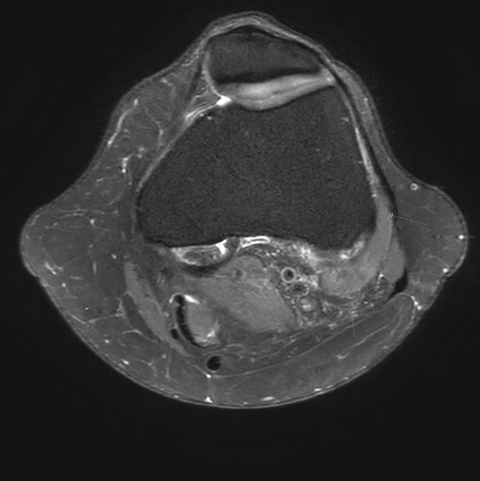
🚀 It's time for Case #107, brought to you by @BIDMC_MSKImg!
➡️ Palpable mass. Top 2 images at presentation. Bottom 2 images are 3 months later.
❓ What is your diagnosis?
📅 Let us know by next Friday, Nov. 25!
#Radiology #MedTwitter #MSKRad #MedEd
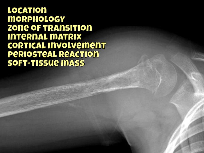
🚀 It's time for Case #104:
➡️ History withheld
❓ What is your diagnosis based on the images provided?
📅 Let us know by next Friday, Oct. 28!
#radiology #MedTwitter #MSKRad #caseoftheweek
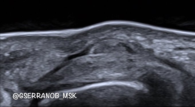
But not very often can we identify the margins of the gap. That's because when we do fist maneuvers, everything tenses up, making the gap challenging to visualize.
With a passive digital mobilization on a relaxed tendon, we can perfectly see the gap.
#mskrad
Case 30 “The Many Faces of Rheumatoid Arthritis”
Three cases in a row!! All you have to do is swipe left. It’s all there 😉
#radres #mskrad #orthotwitter #rheumatoidarthritis
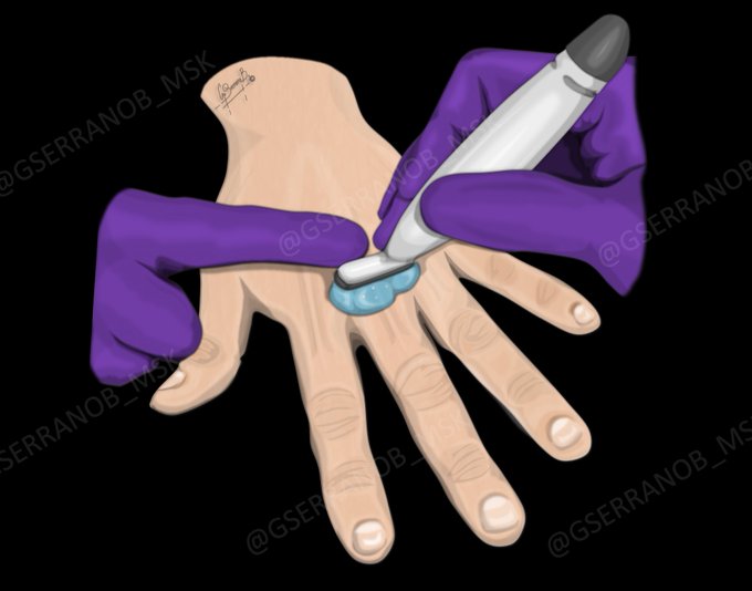
Illustrations and correlation with ultrasound images of the previous video of a full-thickness / full-width laceration of one of the lateral bands in the PIP, but only partial involvement of the mediolateral width of the dorsal apparatus.
#MSKRad
(2/4) As requested by multiple Twitter peeps…here we go with more slides (I’ll post 16 of 35) of our #RSNA21 exhibit:
👍“Thumbs Up Or Down? 👎
MRI of 1st CMC Joint Arthroplasty”
#MSKRad #orthotwitter #radres @ssr_rwg @SSRbone @intskeletal @ESSRmsk @MskSerme @ocad_msk
Illustrations of pronator teres syndrome on ultrasound.
The video case with the dynamic maneuvers I will upload it tomorrow.
The dynamics are a game-changer on the diagnosis.
#medianerve #pronatorteressyndrome #pronatorteres #mskrad #radres
Teenager soccer player, groin pain. Slight separation and traumatic inflammation of traction epiphysis at attachment of sartorius muscle and tensor fascia lata. Edema surounding gluteus minimus origin at ilium. Anterior Superior Iliac Spine Apophysitis. #MSKrad #hip #orthopedics
Illustration of a nice case of sesamoid avulsion of intersesamoid ligament of the thumb. Correlation drawing-ultrasound-MRI (re-posted with a small error correction, sorry).
#mskrad #mskultrasound #radiology #radres #orthotwitter #medart
Patient in her 50ies, chest wall swelling. Sclerosis and osseous hyperostosis of left manubrium sterni, changes located to first sternocostal joint. Osseous proliferation on surface of the bone. No skin or inflammatory disease, no trauma. #MSKrad #orthopaedics #rheumatology (1/3)
Oppenheimer’s ossicle at caudal aspect of right inferior L4 articular process. It might be mistaken for a facet fracture, originates from a horizontal cleft through inferior articular process, shows regular cortical bone in periphery. Learned that from @docskalski here. #MSKrad
Bilateral muscular mass lateral to the distal metaphyseal femur. Anomalous Origin of the Lateral Head of the Gastrocnemius Muscles. https://t.co/0JXiVsObdq #MSKrad #knee #radiology #orthotwitter #FOAMrad #radres
I like saying "I told you so", but don't get to say it nearly enough🤣 For those who mock my reporting of #spine transitional anatomy- you can see the pain here! (Castelvi Ia / IIa)- enhancing articulation (8yrs!) #neurorad #mskrad #radres #Neurosurgery #orthotwitter @docskalski
The current @Radiopaedia featured video is a 7 case Upper Limb X-ray tutorial I recorded during the recent lockdown. Big thanks to my first year registrars for letting the world eavesdrop!
WATCH: https://t.co/HKdZaSa4e3
#foamed #foamrad #radres #mskrad
40yo trauma patient. There’s no injury but there is a syndrome. What is it?
Submit answers as GIF, emoji or haiku ONLY
👤 Case courtesy of @drcraighacking via @Radiopaedia #foamed #foamrad #mskrad #radres
Woman, 40yo
* Parosteal lipoma
Juxtacortical fatty mass with adjacent cortical thickening
- the amount of hyperostosis can vary: small area >>> exuberant exostoses
#mskrad #radiology #radiologia #msk
Young patient with #shoulder instability. No arthrography prescribed. Multiple gleno-humeral intra-articular ossified loose bodies consistent with synovial osteochondromatosis of the shoulder. Secondary to trauma or less likely primary. #MSKrad #orthotwitter #radiology #radres


